

Epub 2022 Jan 31.
The optic nerve head in glaucoma
Affiliations
- PMID: 35210701
- PMCID: PMC8862619
Item in Clipboard
The optic nerve head in glaucoma
Community Eye Health.
2021.
No abstract available
Figures
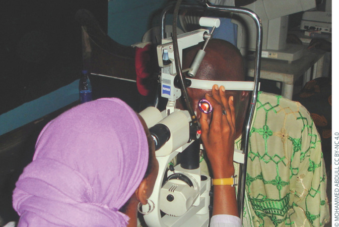
Examining the optic nerve using a slit lamp and posterior pole lens. This offers a stereo view, high magnification, and good illumination and is best performed through a dilated pupil.
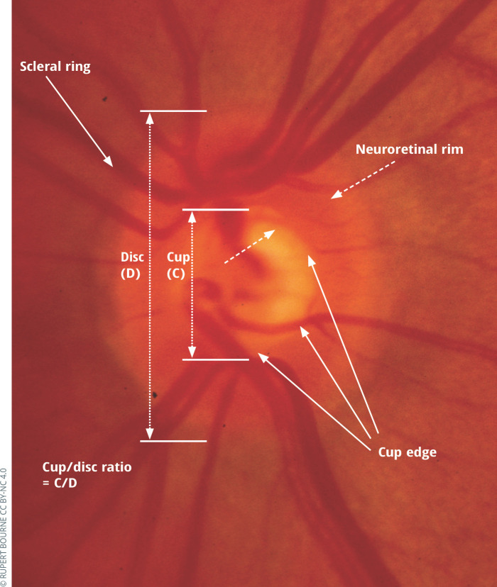
Normal optic nerve head.
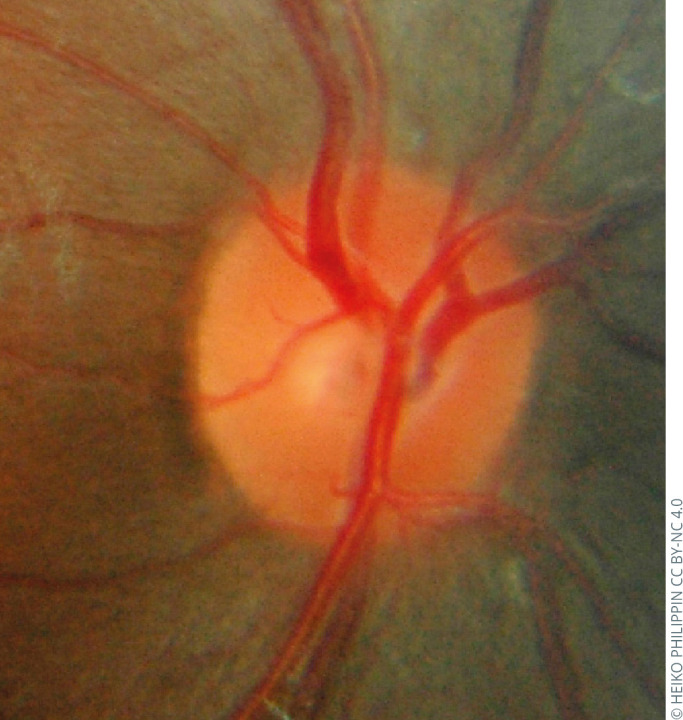
Normal optic nerve head of a young African patient.
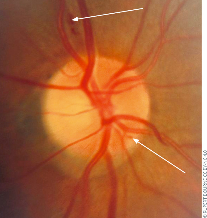
Glaucomatous optic neuropathy: splinter haemorrhages.
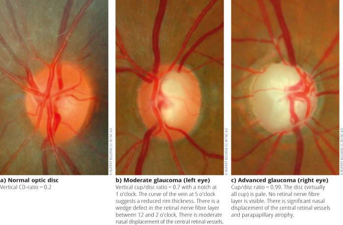
Normal optic disc (a) and glaucomatous optic nerve heads of two patients with different severities of glaucoma.
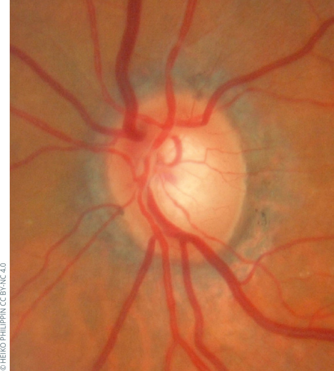
Glaucomatous optic nerve head of a patient with pseudoexfoliation glaucoma (PXFG). The demarcation of the cup by the blood vessels differs from the margin between the pallor of the base of the cup and the surrounding pinker colour between this and the disc edge. Focussing on the colour difference is misleading. One should judge the edge of the rim by the change in direction of the small and medium-sized vessels which, in this case, indicates a thinner rim than might be suspected by the colour difference.
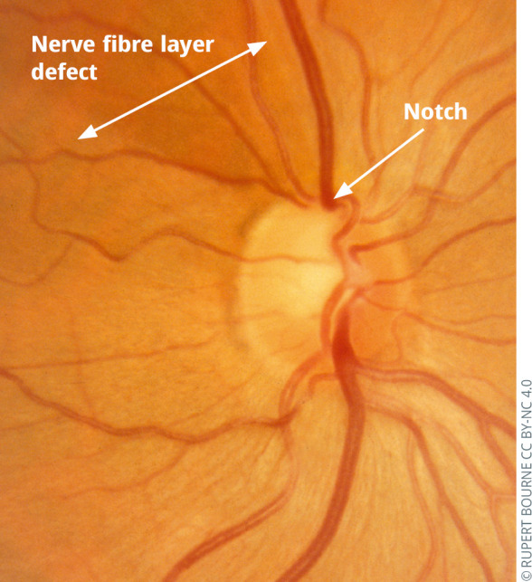
Glaucomatous optic neuropathy: focal enlargement of cup (notch) and nerve fibre layer defect.
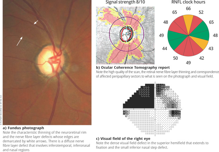
Fundus photograph, ocular coherence tomography report and visual field of the right optic nerve of a patient with glaucoma.
References
-
- Fingeret M, Medeiros FA, Susanna R, Jr, Weinreb RN. Five rules to evaluate the optic disc and retinal nerve fibre layer for glaucoma. Optometry. 2005;76(11):661–8. - PubMed
-
- Iester M, Garway-Heath D, Lemij H. (Eds). Optic nerve head and retinal nerve fibre analysis. Savona, Italy: Dogma Publishing: 2005.
-
- Gupta N, Aung T, Congdon N, Dada T, Lerner F, Olawoye S, et al. ICO Guidelines for Glaucoma Eye Care. Int Counc Ophthalmol. 2015;32(October):1–20.
-
- Sanchez-Galeana C, Bowd C, Blumenthal EZ, Gokhale PA, Zangwill LM, Weinreb RN. Using optical imaging summary data to detect glaucoma. Ophthalmology. 2001. Oct;108(10):1812–8. - PubMed
-
- Li G, Fansi AK, Boivin J-F, Joseph L, Harasymowycz P. Screening for glaucoma in high-risk populations using optical coherence tomography. Ophthalmology. 2010. Mar;117(3):453–61. - PubMed
LinkOut - more resources
Full Text Sources