

Review
doi: 10.1055/s-0041-1740939.
eCollection 2022 Feb.
Embolic Agents: Coils
Affiliations
- PMID: 35210741
- PMCID: PMC8856776
- DOI: 10.1055/s-0041-1740939
Item in Clipboard
Review
Embolic Agents: Coils
Semin Intervent Radiol.
.
No abstract available
Figures
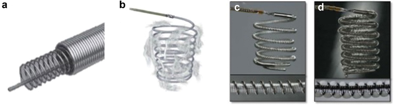
Types of coil materials and coverings. (
a
) Bare platinum coil. (
b
) Fibered helical coil with nylon covering. (
c
) Terumo Azur hydrogel-coated coil preexpansion. (
d
) Hydrogel coil after exposure to blood and expansion of hydrogel covering (∼20 minutes after deployment). (Images used with permissions from Terumo Interventional Systems.)
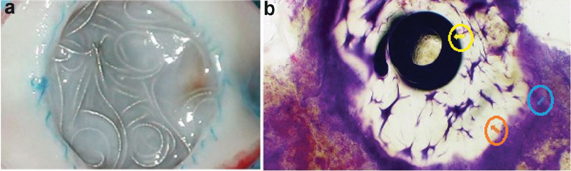
Neointimal proliferation, thrombus formation, and vessel occlusion from coil deployment. (
a
) Gross specimen demonstrating tissue proliferation at the site of hydrogel coil deployment at ∼3 months postintervention. (
b
) Microscopy at 72 days demonstrating neointimal hyperplasia and thrombus formation (blue arrow) adjacent to hydrogel (orange arrow) and platinum coil (yellow arrow). (Images used with permissions from Terumo Interventional Systems.)
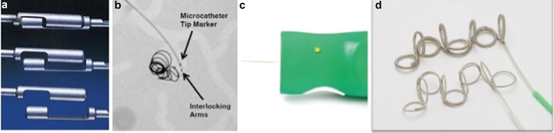
Detachable coil systems. (
a
) Boston scientific detachable interlock and IDC embolization coil via a coupling mechanism. (
b
) Fluoroscopy demonstrating interlocking coil microcatheter and deployed coils. (
c
) Terumo Azur detachable coil remote with coil detachment at push of a button. (
d
) Detachable Azur coils and deployment catheter. (Images used with permissions from Boston Scientific and Terumo Interventional Systems.)
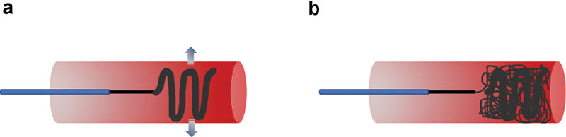
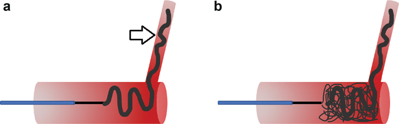
Scaffolding technique for coil deployment. (
a
) An oversized, high radial force (arrows) coil is first deployed into the target vessel to act as a scaffold and to prevent distal coil migration. (
b
) Several softer, smaller coils can then be deployed to achieve complete vessel occlusion.
Anchoring technique for coil deployment. (
a
) A coil is first deployed partially into a side branch (arrow), as well as within the target vessel, acting as an anchor for all subsequent coils. (
b
) Several additional coils can then be densely packed to achieve complete vessel occlusion.
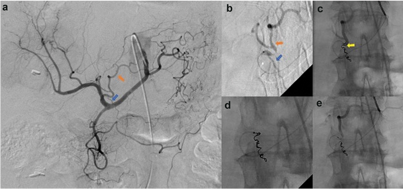
Off-target embolization from catheter reflux and kick-back. (
a
) Celiac artery angiography demonstrates a right gastric artery (blue arrow) arising from the proximal left hepatic artery (orange arrow). Embolization of the right gastric artery is attempted to avoid off-target delivery of radio-embolics to the stomach. (
b
) A microcatheter is used to catheterize the right gastric artery (blue arrow) arising from the left hepatic artery (orange arrow). (
c
) The microcatheter refluxes out of the target right gastric artery with deployment of the last embolization coil (yellow arrow), which appears to be within the left hepatic artery. (
d
) A snare is used to retrieve the unintentional coil deployment within the left hepatic artery. (
e
) Completion angiography demonstrating complete occlusion of the right gastric artery and appropriate contrast-related flow within the left hepatic artery.
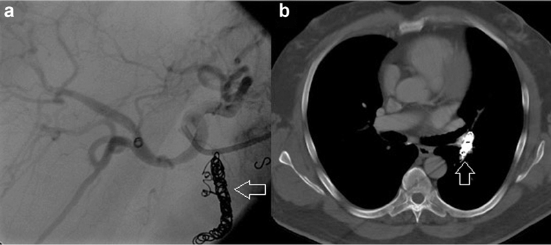
Migration of coil pack. (
a
) Visualization of coil pack in gastroduodenal artery (arrow) pre-radioembolization of the hepatic artery distribution. (
b
) The coil pack (arrow) has migrated to the pulmonary artery.
Similar articles
-
Transvenous embolization of dural carotid cavernous fistulas: the role of liquid embolic agents in association with coils on patient outcomes.J Neurointerv Surg. 2018 May;10(5):461-462. doi: 10.1136/neurintsurg-2017-013318. Epub 2017 Aug 19. J Neurointerv Surg. 2018. PMID: 28823989
-
Relative cost comparison of embolic materials used for treatment of wide-necked intracranial aneurysms.J Neurointerv Surg. 2010 Jun;2(2):163-7. doi: 10.1136/jnis.2009.001719. J Neurointerv Surg. 2010. PMID: 21990601
-
In-vitro thrombogenicity assessment of polymer filament modified and native platinum embolic coils.J Neurol Sci. 2014 Apr 15;339(1-2):97-101. doi: 10.1016/j.jns.2014.01.030. Epub 2014 Jan 31. J Neurol Sci. 2014. PMID: 24553053
-
Evolution of Embolic Agents in Interventional Neuroradiology.Clin Neuroradiol. 2015 Oct;25 Suppl 2:333-9. doi: 10.1007/s00062-015-0419-6. Epub 2015 Jun 18. Clin Neuroradiol. 2015. PMID: 26084977 Review.
-
Embolic agents used for bronchial artery embolisation in massive haemoptysis.Expert Opin Pharmacother. 2004 Feb;5(2):361-7. doi: 10.1517/14656566.5.2.361. Expert Opin Pharmacother. 2004. PMID: 14996632 Review.
Cited by
-
Embolic Agents: Vascular Plugs.Semin Intervent Radiol. 2022 Dec 20;39(5):526-532. doi: 10.1055/s-0042-1758112. eCollection 2022 Oct. Semin Intervent Radiol. 2022. PMID: 36561938 Free PMC article. Review. No abstract available.
-
Endovascular embolization of persistent liver injuries not responding to conservative management: a narrative review.J Trauma Inj. 2023 Sep;36(3):165-171. doi: 10.20408/jti.2023.0040. Epub 2023 Sep 15. J Trauma Inj. 2023. PMID: 39381705 Free PMC article. Review.
-
Enhancing precision in vascular embolization: evaluating the effectiveness of the intentional early detachment technique with detachable coils in complex cases.CVIR Endovasc. 2024 Apr 25;7(1):40. doi: 10.1186/s42155-024-00453-7. CVIR Endovasc. 2024. PMID: 38662076 Free PMC article.
-
Embolic Agents: Particles.Semin Intervent Radiol. 2023 Jul 20;40(3):315-322. doi: 10.1055/s-0043-1769744. eCollection 2023 Jun. Semin Intervent Radiol. 2023. PMID: 37565087 Free PMC article. Review. No abstract available.
-
Improvement in Quality of Life Following Celiprolol Hydrochloride Administration in a Patient with Vascular Ehlers-Danlos Syndrome: A Case Report.Ann Vasc Dis. 2023 Jun 25;16(2):146-149. doi: 10.3400/avd.cr.22-00130. Ann Vasc Dis. 2023. PMID: 37359101 Free PMC article.
References
-
- Mullan S. Experiences with surgical thrombosis of intracranial berry aneurysms and carotid cavernous fistulas. J Neurosurg. 1974;41(06):657–670. - PubMed
-
- Gianturco C, Anderson J H, Wallace S. Mechanical devices for arterial occlusion. Am J Roentgenol Radium Ther Nucl Med. 1975;124(03):428–435. - PubMed
-
- Yuki I, Lee D, Murayama Y. Thrombus organization and healing in an experimental aneurysm model. Part II. The effect of various types of bioactive bioabsorbable polymeric coils. J Neurosurg. 2007;107(01):109–120. - PubMed
-
- Kauvar D S, Schechtman D W, Thomas S B. Endovascular embolization techniques in a novel swine model of fatal uncontrolled solid organ hemorrhage and coagulopathy. Ann Vasc Surg. 2021;70:143–151. - PubMed
