Review
. 2022 Feb 8:13:830292.
doi: 10.3389/fimmu.2022.830292. eCollection 2022.

Advances in CAR-T Cell Genetic Engineering Strategies to Overcome Hurdles in Solid Tumors Treatment

Affiliations
Review

Advances in CAR-T Cell Genetic Engineering Strategies to Overcome Hurdles in Solid Tumors Treatment

Alain E Andrea et al. Front Immunol. .

Abstract

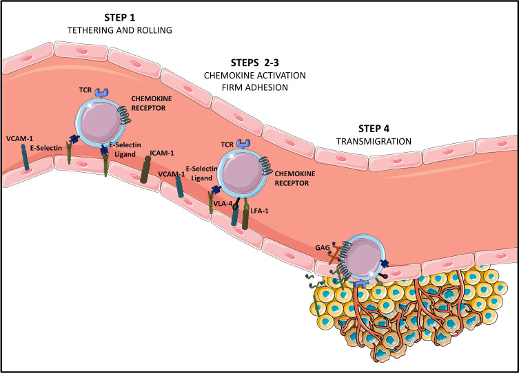
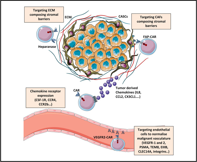
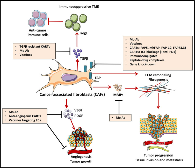
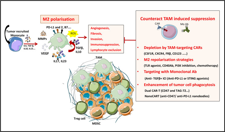
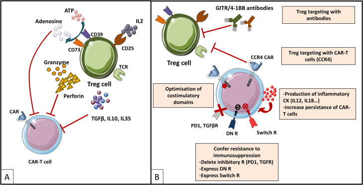
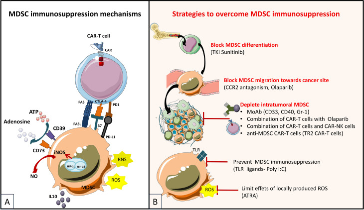
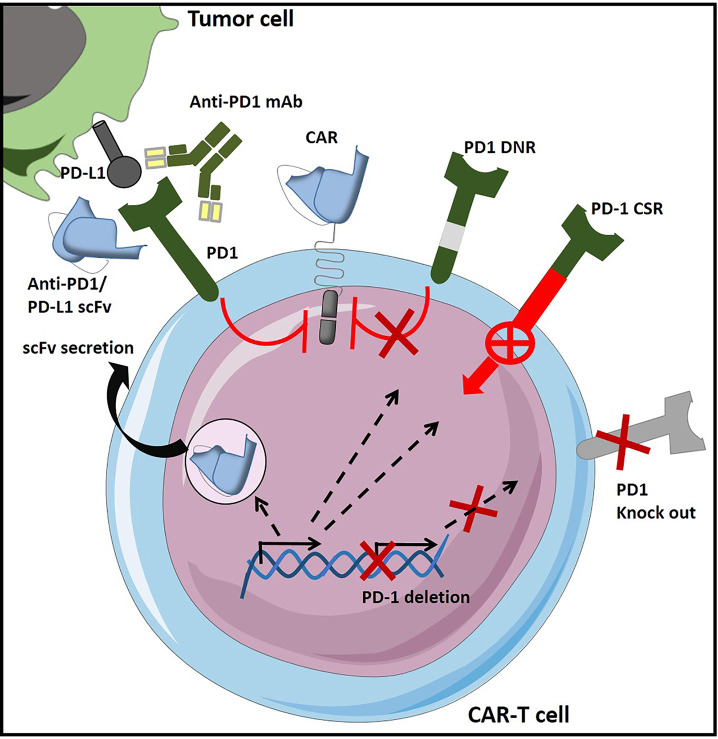
During this last decade, adoptive transfer of T lymphocytes genetically modified to express chimeric antigen receptors (CARs) emerged as a valuable therapeutic strategy in hematological cancers. However, this immunotherapy has demonstrated limited efficacy in solid tumors. The main obstacle encountered by CAR-T cells in solid malignancies is the immunosuppressive tumor microenvironment (TME). The TME impedes tumor trafficking and penetration of T lymphocytes and installs an immunosuppressive milieu by producing suppressive soluble factors and by overexpressing negative immune checkpoints. In order to overcome these hurdles, new CAR-T cells engineering strategies were designed, to potentiate tumor recognition and infiltration and anti-cancer activity in the hostile TME. In this review, we provide an overview of the major mechanisms used by tumor cells to evade immune defenses and we critically expose the most optimistic engineering strategies to make CAR-T cell therapy a solid option for solid tumors.

Keywords: Angiogenesis; CAR-T cell immunotherapy; Chemokines; Immune checkpoints; Solid tumor; Tumor Homing; Tumor microenvironment; Tumor stroma.


Conflict of interest statement

The authors declare that the research was conducted in the absence of any commercial or financial relationships that could be construed as a potential conflict of interest.

Figures

Figure 1
Figure 1
Steps of T cell homing to tumor tissues [Adapted from Sackstein et al. (11)]. Tumor infiltrating CD8+ effector T lymphocytes (Teffs) presenting a specific tumor antigen circulate in the blood stream. They express homing molecules allowing for their oriented migration towards the tumor (like CXCR3 and CCR5-chemokine receptors), as well as ligands allowing binding to endothelial cells (E-selectin ligands and VLA-4 and LFA-1 integrins at suboptimal levels). Circulating Teffs tether and roll on the endothelium (STEP 1) via engagement of E-Selectin ligands with endothelial E-Selectin, which slows down Teffs, and allows firm adhesion to the endothelium (STEP 2). In this second step of Teffs entry into tumoral tissues, chemokines produced by cancer cells or by stromal cells from the TME (CXCL9, CXCL10, CCL5…) bind chemokine receptors. This binding of chemokine receptors to their ligands elicits activation of VLA-4 and LFA-1, allowing for VLA-4/VCAM-1 and LFA-1/ICAM-1 firm adhesion (STEP 3). Firmly adherent Teffs undergo transendothelial migration (STEP 4), to infiltrate the TME and establish cell-to-cell contact with tumor cells, via TCR-based recognition of cancer antigens presented on HLA molecules.
Figure 2
Figure 2
Strategies enhancing tumor trafficking and penetration [Adapted from Rafiq et al. (31)]. The trafficking of CAR-T cells towards tumor sites can be enhanced by engineering CAR-T cells expressing chemokine receptors (as for example CSF-1R, CCR4 or CCR2b) specific for tumor-derived chemokine ligands (IL8, CCL2, CXCL1…). Tumor penetration of CAR-T cells can be enhanced by various strategies: (1) normalizing the malignant vasculature by targeting tumor blood vessels via CAR targeting of endothelial/tumoral antigens (like VEGFR, EIIIB, TEM8, integrins.), and (2) targeting physical barriers in the tumor microenvironment (TME) like the extracellular matrix (ECM) or the cancer associated fibroblasts (CAFs).
Figure 3
Figure 3
Strategies to counteract protumorigenic effects of CAFs [Adapted from Kakarla et al. (148)]. Cancer associated fibroblasts (CAF)-directed anti-cancer therapies are one of the weapons of tumor targeting which is directed against the stromal compartment. Strategies depicted in this figure aim at inhibiting cancer associated fibroblasts (CAFs) functions and are based on targeting crucial signals and effectors of CAFs such as cytokines (TGFβ) and growth factor pathways (VEGF, PDGF…). For instance, CAF-derived extracellular matrix proteins (MMPs) and associated signaling can be targeted with monoclonal antibodies (MAb), to induce stromal depletion and increase immune T cell infiltration. Blocking some targets like TGFβ, can act both upstream and downstream, by blocking CAF formation and attenuating downstream signaling in CAFs that are already established. FAP targeting aims at blocking CAFs ability to exert tumor promoting effects in the TME. Targeting FAP can be done by using either MAb/antibody-drug conjugates, immunoconjugates or peptide-drug complexes, FAP-specific CAR-T cells or strategies of gene-knock out. Some other strategies, not depicted in this figure aim at CAFs direct depletion or CAFs normalization towards an inactive phenotype.
Figure 4
Figure 4
Strategies to overcome TAM’s induced suppression in the TME. TAMs are a tumor promoting immune populations derived under a specific cytokine milieu either from blood circulating monocytes or from tumor resident macrophages. TAMs exert their tumor promoting and immunosuppressive role by means of cell-to cell contact (inhibitory check point ligands), by secreting soluble factors (like cytokines IL10, IL17, L23), by producing ECM-modifying enzymes (MMPs) or by producing reactive species of oxygen (ROS). All these factors promote tumor progression. TAMs directed therapies in the TME aim either at (1) specifically depleting the TAM population, at (2) reprogramming M2 towards proinflammatory M1 phenotypes, at (3) targeting TAM-secreted factors or 4) at enhancing TAM’s phagocytic functions in the TME.
Figure 5
Figure 5
Immunosuppressive mechanisms exerted by Tregs in the TME (A) and engineering strategies to surmount Treg-induced immunosuppression (B) [Adapted from Togashi et al. (212), and Rodriguez-Garcia et al. (213)]. (A) depicts mechanisms for regulatory T (Treg) cells immunosuppressive effects on CAR-T cells based on their physiologic roles. Tregs are immunosuppressive cells highly dependent on IL-2. They bind to and deplete IL-2 from their surroundings, thus reducing availability to effector T (Teff) cells by constitutively expressing the high affinity IL-2 receptor (IL2R) subunit-α (CD25). Treg cells also produce immunosuppressive cytokines (IL-10, IL-35 and TGFβ), which can downregulate the activity of both Teffs and antigen presenting cells (APCs) and they exert direct cytotoxic effects by secreting granzymes and perforin. Moreover, Treg cells release large amounts of ATP, which is converted to adenosine (by CD39 and CD73) that can provide immunosuppressive signals to Teff cells and APCs. Other indirect mechanisms not depicted in the figure by which Tregs exert immunosuppressive effects are mediated by APC, as for instance Tregs expression of cytotoxic T lymphocyte antigen 4 (CTLA-4), which binds to CD80/CD86 on APCs, thereby transmitting suppressive signals to these cells and reducing their capacity to activate Teff cells. (B) shows therapeutic strategies to overcome the immunosuppressive TME sustained by Tregs. Some strategies are based on elimination of Tregs by CAR-T cells or combinations of CAR-T cells with monoclonal antibodies (mAbs) or drugs. CAR-T cells have been designed to target antigens expressed by Tregs for direct depletion. Other strategies are based on immunomodulation of the TME in order to increase CAR-T cells performance: 1) expression of proinflammatory cytokines by CAR-T cells and 2) optimization of costimulatory signaling domains in order to reduce IL-2 secretion and impair Treg expansion and tumor infiltration. Last type of strategies are meant to confer an intrinsic resistance to immunosuppression to CAR-T cells, either by endowing them with 1) dominant-negative receptors (DN R) meant to disrupt signaling, or 2) a chimeric switch receptor (CSR or Switch R) to convert negative signaling into a positive one, or by abrogating the expression of inhibitory receptors (like PD1 of TGFβ receptors) using genome-editing tools (knock out).
Figure 6
Figure 6
Immunosuppressive mechanisms exerted by MDSCs in the tumor microenvironment (A) and engineering strategies to surmount MDSC-induced immunosuppression (B) [Adapted from Krishnamoorthy et al. (250)]. (A) MDSC exert immunosuppressive effects in the TME (tumor microenvironment) by secretion of IL10 (which activates other immunosuppressive cells such as Tregs). Moreover, MSDCs can induce upregulation of checkpoint molecules (CTLA4, PD1) on T-cells, further inducing T-cell anergy, or can upregulate Fas which induces T-cell apoptosis by contact Fas/Fas-L mechanism. As an effect of hypoxia in the TME, MDSC can contribute to adenosine production by upregulation of CD73 and CD39. MDSCs also produce reactive oxygen (ROS) and nitrogen (RNS) species that can decrease T-cell proliferation and alter antigen recognition capabilities. (B) Strategies for targeting MDSCs in cancer are for example the prevention of MDSC differentiation from hematopoietic stem cells by the usage of Sunitinib, a tyrosine kinase inhibitor (TKI) that inhibits crucial factors for MDSC differentiation (VEGF and STAT3 activity). Second type of strategy is to prevent MDCS migrating to the tumor by targeting chemokine/chemokine receptor axes (CCR2/CCL2). Third, MDSCs depletion from the tumor can be achieved by using immunotherapy (depleting antibodies targeting CD33/gemtuzumab ozogamicin (GO), CD40 or Gr1) or chemotherapy. And least, mitigating the immunosuppressive effects of MDSCs at the tumor site can be realized by reducing the local effects of ROS (with ATRA or all-trans-retinoic acid) or by using TLR stimulation with specific ligands (TLR3 ligand polyinosinic-polycytidylic acid Poly I:C)
Figure 7
Figure 7
CAR-T cell engineering strategies to overcome inhibition form negative immune checkpoint regulation – Example of PD1/PD-L1 axis targeting in CAR-T cells [Adapted from Rafiq et al. (31)]. In order to prevent CAR-T cell exhaustion and immunosuppression in the TME, different strategies can be used, like combination of CAR-T cells with immune checkpoint inhibitors (ICIs like anti-PD1 or PD-L1 antibodies). PD1-medited inhibition can also be surmounted by designing CAR-T cells that secrete either PD-1-blocking or PD-L1 blocking scFv. Other means of shielding CAR-T cells from the inhibitory effect of the PD1/PD-L1 interaction is to design genetically modified CAR-T cells that express a dominant negative PD-1 receptor (PD-1 DNR) which interferes with PD1 downstream signaling or a PD-1 chimeric switch receptor (CSR), which converts an inhibitory signaling into an activating one. Last type of strategy is based on PD1 expression deletion either by genetic knock-out or by means of shRNA (short hairpin RNA) inhibition.

Similar articles

Cited by

References

    1. Zhang C, Liu J, Zhong JF, Zhang X. Engineering CAR-T Cells. biomark Res (2017) 5:22. doi: 10.1186/s40364-017-0102-y - DOI - PMC - PubMed
    1. Chmielewski M, Abken H. TRUCKS, the Fourth-Generation CAR T Cells: Current Developments and Clinical Translation. Adv IN Cell AND Gene Ther (2020) 3:e84. doi: 10.1002/acg2.84 - DOI
    1. Fridman WH, Pagès F, Sautès-Fridman C, Galon J. The Immune Contexture in Human Tumours: Impact on Clinical Outcome. Nat Rev Cancer (2012) 12:298–306. doi: 10.1038/nrc3245 - DOI - PubMed
    1. Mantovani A, Marchesi F, Malesci A, Laghi L, Allavena P. Tumour-Associated Macrophages as Treatment Targets in Oncology. Nat Rev Clin Oncol (2017) 14:399–416. doi: 10.1038/nrclinonc.2016.217 - DOI - PMC - PubMed
    1. Chow MT, Luster AD. Chemokines in Cancer. Cancer Immunol Res (2014) 2:1125–31. doi: 10.1158/2326-6066.CIR-14-0160 - DOI - PMC - PubMed

Substances