. 2022 Feb 21;10(1):coac004.
doi: 10.1093/conphys/coac004. eCollection 2022 Jan 1.

Isolation and characterization mesenchymal stem cells from red panda (Ailurus fulgens styani) endometrium

Affiliations

Isolation and characterization mesenchymal stem cells from red panda (Ailurus fulgens styani) endometrium

Dong-Hui Wang et al. Conserv Physiol. .

Abstract

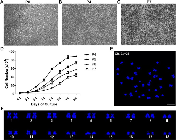
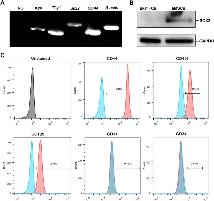
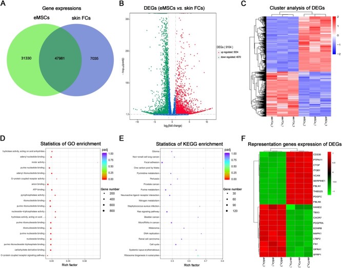
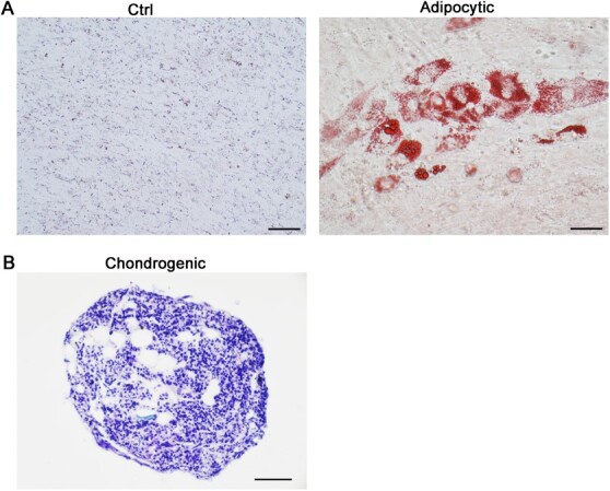
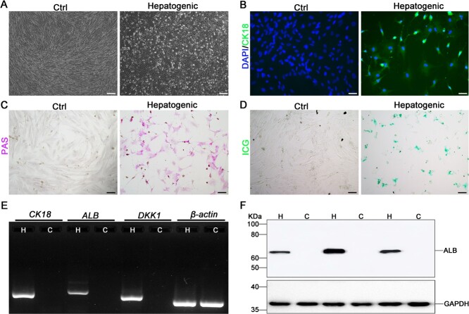
Endometrial mesenchymal stem cells (eMSCs) are undifferentiated endometrial cells with self-renewal, multidirectional differentiation and high proliferation potential. Nowadays, eMSCs have been found in a few species, but it has never been reported in endangered wild animals, especially the red panda. In this study, we successfully isolated and characterized the eMSCs derived from red panda. Red panda eMSCs were fibroblast-like, had a strong proliferative potential and a stable chromosome number. Pluripotency genes including Klf4, Sox2 and Thy1 were highly expressed in eMSCs. Besides, cultured eMSCs were positive for MSC markers CD44, CD49f and CD105 and negative for endothelial cell marker CD31 and haematopoietic cell marker CD34. Moreover, no reference RNA-seq was used to analyse the eMSCs transcriptional expression profile and key pathways. Compared with skin fibroblast cell group, 9104 differentially expressed genes (DEGs) were identified, among which are 5034 genes upregulated, 4070 genes downregulated and the top 20 enrichment pathways of DEGs in Gene Ontology (GO) and the Kyoto Encyclopedia of Genes Genomes (KEGG) mainly associated with G-protein coupled receptor signalling pathway, carbohydrate derivative binding, nucleoside binding, ribosome biogenesis, cell cycle, DNA replication, Ras signalling pathway and purine metabolism. Among the DEGs, some representative genes about promoting MSCs differentiation and proliferation were upregulated and promoting fibroblasts proliferation were downregulated in eMSCs group. Red panda eMSCs also had multiple differentiation ability and could differentiate into adipocytes, chondrocytes and hepatocytes. In conclusion, we, for the first time, isolated and characterized the red panda eMSCs with ability of multiplication and multilineage differentiation in vitro. The new multipotential stem cell could be beneficial not only for the germ plasm resources conservation of red panda, but also for basic or pre-clinical studies in the future.

Keywords: Cell culture; cell differentiation; endometrium; mesenchymal stem cell; red panda.


Figures

Figure 1
Figure 1
Morphological characteristics, growth curves and chromosome spreads of red panda eMSCs. (A) Representative image of primary red panda eMSCs after 9 days culture. The primary cells were fibroblast like and epithelial like, presenting triangular, fusiform, ovoid or polygonal shapes. Scale bar, 200 μm. (B) Representative image of red panda eMSCs at passage 4. Fibroblast-like shape and presented polygonal or long spindle shape. Scale bar, 100 μm. (C) Representative image of red panda eMSCs at passage 7. Cells still maintain fibroblast-like and long spindle shapes. Scale bar, 100 μm. (D) The growth curves of the cells from passages 4–7. Quantified data show the mean ± SEM. (E) Chromosome spreads of red panda eMSCs at passage 8. Scale bar, 10 μm. (F) Analysis of the chromosome spreads of (E). The correct number of chromosomes in red panda is 2n = 36.
Figure 2
Figure 2
Characterization of red panda eMSCs. (A) RT-PCR analysis of pluripotency genes in eMSCs. β-actin was used as a control for RNA sample quality. (B) The expression of SOX2 was detected in red panda eMSCs and skin FCs by western blot. GAPDH was used as the loading control. (C) Red panda eMSCs were stained with phycoerythrin (PE)-conjugated CD44, CD49f, CD105, CD34 or PE-Cyanine7 conjugated CD31 antibodies. Blue areas, signal from isotype controls; red areas, signal from the specific cell surface marker; grey areas, unstained black control.
Figure 3
Figure 3
Differential gene expression analysis between red panda eMSCs and skin FCs. (A) The Venn diagram of the number of predicted genes in red panda eMSCs and skin FCs. (B) Volcano map of DEGs between red panda eMSCs and skin FCs. The x-axis is the log2 scale of the fold change of gene expression in eMSCs and skin FCs (log2(fold change)). Negative values indicate downregulation; positive values indicate upregulation. The y-axis is the minus log10 scale of the adjusted P-values (−log10(padj)), which indicate the significant level of expression difference. The red dots represent significantly upregulated genes with at least 2-fold change, while the green dots represent significantly downregulated genes with at least 2-fold change. (C) Heatmap of the differentially expressed genes between red panda eMSCs and skin FCs. Red stripes represent high expression genes, while blue stripes represent low expression genes. (D) Top 20 significantly enriched GO terms. (E) Top 20 enriched KEGG pathways. (F) Heatmap of the representative functional genes expression of DEGs between red panda eMSCs and skin FCs. Red stripes represent high expression, while green stripes represent low expression.
Figure 4
Figure 4
Adipocytic and chondrogenic differentiation of red panda eMSCs. (A) Red panda eMSCs were cultured in the adipocytic induction medium for 8 days, and then evaluated by Oil-red O staining to reveal lipid vacuoles. Untreated cells were used as control. Scale bar, 50 μm. (B) Red panda eMSCs were cultured in the chondrogenic differentiation medium for 21 days, and then evaluated by toluidine blue staining to reveal the sulfated proteoglycans of the cartilage matrices. Scale bar, 50 μm.
Figure 5
Figure 5
Hepatogenic differentiation of red panda eMSCs. (A) Bright field images of red panda eMSCs cultured in hepatogenic differentiation medium for 16 days, and eMSCs were changed to flat polygonal morphology. Untreated cells were used as control. Scale bar, 200 μm. (B) Red panda eMSCs after hepatogenic differentiation were stained with anti-CK18 (hepatogenic marker, green), and the nucleus were stained with DAPI (blue). Untreated cells were used as control. Scale bar, 50 μm. (C) Red panda eMSCs after hepatogenic differentiation were stained with PAS staining (purple signal). Untreated cells were used as control. Scale bar, 100 μm. (D) Red panda eMSCs after hepatogenic differentiation were detected by ICG uptake (green signal). Untreated cells were used as control. Scale bar, 100 μm. (E) The expressions of liver-specific genes in hepatogenic-induced (H) and control (C) red panda eMSCs were detected by using RT-PCR. β-actin was used as a reference gene. (F) The expressions of ALB in hepatogenic-induced (H) and control (C) red panda eMSCs were detected by using western blot. GAPDH was used as the loading control.

Similar articles

Cited by

References

    1. Alifi F, Asgari HR (2020) Alteration in expression of primordial germ cell (PGC) markers during induction of human amniotic mesenchymal stem cells (hAMSCs). J Reprod Infertil 21: 59–64. - PMC - PubMed
    1. An JH, Li FP, He P, Chen JS, Cai ZG, Liu SR, Yue CJ, Liu YL, Hou R (2020) Characteristics of mesenchymal stem cells isolated from the bone marrow of red pandas. Zoology (Jena) 140: 125775. - PubMed
    1. Bartoletti G, Dong C, Umar M, He F (2020) Pdgfra regulates multipotent cell differentiation towards chondrocytes via inhibiting Wnt9a/beta-catenin pathway during chondrocranial cartilage development. Dev Biol 466: 36–46. - PMC - PubMed
    1. Baszanowska W, Misiura M, Oscilowska I, Palka J, Miltyk W (2021) Extracellular prolidase (PEPD) induces anabolic processes through EGFR, beta1-integrin, and IGF-1R signaling pathways in an experimental model of wounded fibroblasts. Int J Mol Sci 22: 942. - PMC - PubMed
    1. Bian Y, Du Y, Wang R, Chen N, Du X, Wang Y, Yuan H (2019) A comparative study of HAMSCs/HBMSCs transwell and mixed coculture systems. IUBMB Life 71: 1048–1055. - PubMed