. 2022 Feb 15:9:83-98.
doi: 10.2147/JHC.S349966. eCollection 2022.

SLC39A1 Overexpression is Associated with Immune Infiltration in Hepatocellular Carcinoma and Promotes Its Malignant Progression

Affiliations

SLC39A1 Overexpression is Associated with Immune Infiltration in Hepatocellular Carcinoma and Promotes Its Malignant Progression

Xiaowu Ma et al. J Hepatocell Carcinoma. .

Abstract

Background: Solute carrier family 39 member 1 (SLC39A1) has been identified as a zinc ion transport protein that possesses oncogenic properties in various types of cancers. However, its potential function in hepatocellular carcinoma (HCC) remains unknown. This study aimed to investigate the expression profile and potential mechanisms of SLC39A1 in HCC.

Methods: SLC39A1 expression was analyzed using multiple databases. The clinical significance and associated biological pathways of SLC39A1 were investigated using bioinformatics analysis. Potential correlations between SLC39A1 expression and tumor immunity in HCC were also evaluated using single-sample gene set enrichment analysis (GSEA). Sixty paired HCC samples were used to verify the expression pattern of SLC39A1. In vitro studies were performed to investigate the oncogenic effects of SLC39A1 in HCC. Western blot analysis was conducted to further investigate the possible involved signaling pathways.

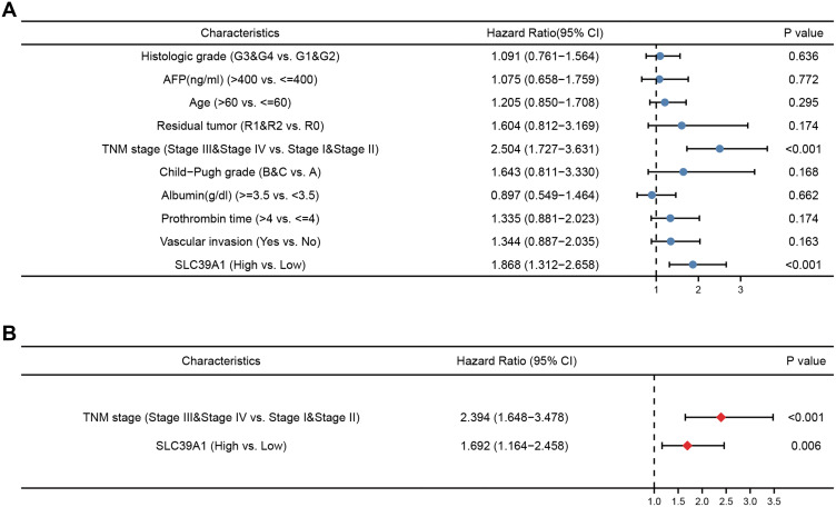
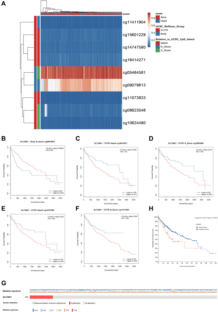
Results: The overexpression of SLC39A1 in HCC was determined by bioinformatics analysis and was confirmed in tissues from our center. SLC39A1 overexpression was also significantly correlated with worse prognosis, advanced TNM stage, and histological grade. GSEA analysis demonstrated that SLC39A1 overexpression was involved in various tumor-related pathways, such as the cell cycle and Wnt signaling pathway. SLC39A1 knockdown repressed the proliferation, invasion, and migration abilities of HCC cells. Furthermore, SLC39A1 knockdown decreased the expression of the tumor progression-related proteins (eg, cyclin D1 and MMP2) and Wnt signaling pathway-related proteins (eg, Wnt3A and β-catenin). In addition, SLC39A1 overexpression may be associated with impaired tumor immunity in HCC, as evidenced by the increased infiltration of Th2 cells and reduced infiltration of cytotoxic cells.

Conclusion: These findings preliminarily suggested the crucial effect of SLC39A1 overexpression on HCC tumor progression and immunosuppression, suggesting its potential as a novel prognostic and therapeutic target in HCC.

Keywords: SLC39A1; Wnt signaling pathway; hepatocellular carcinoma; immune infiltration; prognosis.


Conflict of interest statement

All authors declare that they have no conflicts of interest for this work.

Figures

Figure 1
Figure 1
SLC39A1 is over-expressed in Hepatocellular Carcinoma (HCC). (A) The different expression of SLC39A family between normal and tumor in HCC from Oncomine database. (B) Dynamic expression of SLC39A1 in pan-cancer. (C) Overexpression of SLC39A1 in HCC was identified in HCCDB database. *P < 0.05, **P < 0.01, ***P < 0.001.
Figure 2
Figure 2
Overexpression of SLC39A1 correlates with poor survival and unfavorable clinicopathological characteristics in Hepatocellular Carcinoma (HCC). (AC) SLC39A1 expression of HCC was significantly associated with TNM stage, Histologic grade, and alpha fetoprotein (AFP) level. (DF) Kaplan-Meier analysis showed HCC patients with high expression of SLC39A1 had poor overall survival (D), progression-free survival (E) and disease specific survival (F) than those with low expression in TCGA. (G) ROC (Receiver operator characteristic curve) analysis showed that SLC39A1 was a reliable prognostic predictor in patients with HCC. *P < 0.05, ***P < 0.001.
Figure 3
Figure 3
Univariate and multivariate regression cox analysis of SLC39A1. (A) Univariate cox regression analysis was performed to confirmed high expression of SLC39A1 was associated with overall survival (OS) of HCC patients. (B) Multivariate cox regression analysis was performed to confirmed high expression of SLC39A1 was independent risk factor for OS of HCC patients.
Figure 4
Figure 4
Prognostic value of SLC39A1 DNA methylation and genetic alteration in HCC. (A) The DNA methylation clustered expression of SLC39A1. (BF) Overall survival (OS) curve of cg09079613 (B), cg16414271 (C), cg10624480 (D), cg14747580 (E) and cg11411904 (F) in HCC patients. (G) The overview of genetic alteration in SLC39A1. (H) OS curve between altered and unaltered group of SLC39A1.
Figure 5
Figure 5
Gene Ontology (GO) enrichment analysis and Gene set enrichment analysis (GSEA) of SLC39A1 in HCC. (A) GO enrichment analysis of SLC39A1 related genes in HCC. (BG) GSEA showed that the group with high expression of SLC39A1 was significantly associated with many tumor progression-related pathways, such as Wnt signaling pathway (B), Cell cycle (C), DNA replication (D), TGF-β signaling pathway (E), NOD like receptor signaling pathway (F) and MAPK signaling pathway (G).
Figure 6
Figure 6
The expression of SLC39A1 was associated with the immune infiltration in the tumor microenvironment. (A) Correlation between the relative abundance of 24 immune cells and SLC39A1 expression. (BE) Box plot and correlation diagrams showing the difference of Th2 and cytotoxic cells infiltration level between SLC39A1-high and low group. ***P < 0.001.
Figure 7
Figure 7
The overexpression of SLC39A1 in HCC patients. (A) The differences in the expression of SLC39A1 in 60 tumor and paired normal tissues. (B) The Fold Changes (FC) of SLC39A1 expression in 60 HCC patients. (C) Distribution of three groups (High, Low and No change) in 60 HCC patients. (D) The SLC39A1 expression results of genomic sequence in 20 HCC patients. ****P < 0.0001.
Figure 8
Figure 8
Effects of SLC39A1 on HCC proliferation and metastasis. (A) The change of SLC39A1 expression after transfected sh-SLC39A1. (B and C) CCK-8 assay and EdU assay demonstrated that transfection with sh-SLC39A1 plasmid inhibited the proliferation of Huh7 cell. Scale bar = 100 µm. (D) Transwell assays indicated that migration and invasion ability of Huh7 cell was reduced after transfecting with sh-SLC39A1 plasmid. Scale bar = 100 µm. (E) The downregulation of N-cadherin, MMP2 and CyclinD1 in Huh7 cell transfected with sh-SLC39A1 plasmid was detected by Western blot. (F) The downregulation of Wnt3A, p-GSK3β and β-catenin in Huh7 cell transfected with sh-SLC39A1 plasmid was detected by Western blot. *P < 0.05, **P < 0.01, ***P < 0.001, ****P < 0.0001.

References

    1. Sung H, Ferlay J, Siegel RL, et al. Global cancer statistics 2020: GLOBOCAN estimates of incidence and mortality worldwide for 36 cancers in 185 countries. CA Cancer J Clin. 2021;71(3):209–249. doi:10.3322/caac.21660 - DOI - PubMed
    1. Villanueva A, Longo DL. Hepatocellular carcinoma. N Engl J Med. 2019;380(15):1450–1462. doi:10.1056/NEJMra1713263 - DOI - PubMed
    1. Finn RS, Qin S, Ikeda M, et al. Atezolizumab plus bevacizumab in unresectable hepatocellular carcinoma. N Engl J Med. 2020;382(20):1894–1905. doi:10.1056/NEJMoa1915745 - DOI - PubMed
    1. Ren Z, Xu J, Bai Y, et al. Sintilimab plus a bevacizumab biosimilar (IBI305) versus sorafenib in unresectable hepatocellular carcinoma (ORIENT-32): a randomised, open-label, phase 2–3 study. Lancet Oncol. 2021;22(7):977–990. doi:10.1016/s1470-2045(21)00252-7 - DOI - PubMed
    1. Giraud J, Chalopin D, Blanc JF, Saleh M. Hepatocellular carcinoma immune landscape and the potential of immunotherapies. Front Immunol. 2021;12:655697. doi:10.3389/fimmu.2021.655697 - DOI - PMC - PubMed