Review
. 2022 Feb 8:8:768649.
doi: 10.3389/fmed.2021.768649. eCollection 2021.

Supraspinal Mechanisms Underlying Ocular Pain

Affiliations
Review

Supraspinal Mechanisms Underlying Ocular Pain

Nicholas J Pondelis et al. Front Med (Lausanne). .

Abstract

Supraspinal mechanisms of pain are increasingly understood to underlie neuropathic ocular conditions previously thought to be exclusively peripheral in nature. Isolating individual causes of centralized chronic conditions and differentiating them is critical to understanding the mechanisms underlying neuropathic eye pain and ultimately its treatment. Though few functional imaging studies have focused on the eye as an end-organ for the transduction of noxious stimuli, the brain networks related to pain processing have been extensively studied with functional neuroimaging over the past 20 years. This article will review the supraspinal mechanisms that underlie pain as they relate to the eye.

Keywords: brain; brainstem; eye; fMRI; neuroimaging; ocular; pain; supraspinal.


Conflict of interest statement

The authors declare that the research was conducted in the absence of any commercial or financial relationships that could be construed as a potential conflict of interest.

Figures

Figure 1
Figure 1
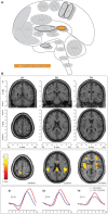
Nociceptive pathways. (A) The path of afferent signal transmission from the periphery to the cortex through major projections of the trigeminothalamic pathway. Reprinted/adapted by permission from Springer Nature Customer Service Centre GmbH: Springer Nature Trigeminothalamic Tract Projections. In: Schmidt R., Willis W. (eds) Encyclopedia of Pain by Ke Ren, Copyright (2007). DOI: https://doi.org/10.1007/978-3-642-28753-4_4626. (B) Decreased brainstem fMRI activity, including PB, during endogenous analgesia. Red/yellow indicates regions where fMRI responses to noxious stimuli demonstrated a signal decrease following conditioned pain modulation. Decreased activation was noted in the SRD, SpVc, and the trigeminal nerve along with the PB. On the left, myelin-stained sections are displayed alongside corresponding MRI images of the brainstem. Reprinted from NeuroImage, Vol 124(Part A), AM Youssef, VG Macefield, LA Henderson, Pain inhibits pain; human brainstem mechanisms, p54–62, Copyright (2020), with permission from Elsevier. DOI: https://doi.org/10.1016/j.neuroimage.2015.08.060. PrV, principal sensory nucleus; SpV, spinal trigeminal nucleus; Vo, subnuclei oralis; Vi, subnucleus interpolaris; Vc, subnucleus caudalis; PB, parabrachial nucleus; SRD, subnucleus reticularis dorsalis; SpVc, spinal trigeminal nucleus caudalis; V, trigeminal nerve; Compas: S, superior; I, inferior; R, right; L, left.
Figure 2
Figure 2
Pain-related areas in the brain and brainstem. (A) A meta-analysis of pain neuroimaging studies defines a set of brain regions consistently active across 222 experiments from 200 reports, including bilateral activity in the secondary somatosensory cortex, insular cortex, midcingulate cortex, and thalamus. Voxel values increase from 1 to 15 with increasing convergence across 15 total main effects meta-analyses that each reflect pain-related activation. Reprinted from Neuroscience & Biobehavioral Reviews, Vol 112, A Xu, B Larsen, EB Baller, JC Scott, V Sharma, A Adebimpe, AI Basbaum, RH Dworkin, RR Edwards, CJ Woolf, SB Eickhoff, CR Eickhoff, TD Satterthwaite, Convergent neural representations of experimentally-induced acute pain in healthy volunteers: A large-scale fMRI meta-analysis, p300–23, Copyright (2020), with permission from Elsevier. https://doi.org/10.1016/j.neubiorev.2020.01.004. (B) Schematic of brain areas related to the processing of the multidimensional experience of pain. Each region is color coded to correspond to its hypothesized dimension of pain, while hatch-marks indicate processing associated with pain-related movement. Thick black borders indicate regions located more lateral to the midline. Relative size of each region is roughly proportional for structures larger than SII. (C) Attention to different features of a painful stimulus can shift activation patterns. Focusing on the unpleasantness of pain vs its location results in different patterns of brain activation when examined by PET, providing evidence that the unique dimensions of pain may be processed in separate brain areas. Reprinted from the European Journal of Neuroscience, Vol 21(11), B Kulkarni, DE Bentley, R Elliott, P Youell, A Watson, SW Derbyshire, RS Frackowiak, KJ Friston, AK Jones, Attention to pain localization and unpleasantness discriminates the functions of the medial and lateral pain systems, p3133-42, Copyright (2005), with permission from John Wiley and Sons. DOI: https://doi.org/10.1111/j.1460-9568.2005.04098.x. SI, primary somatosensory cortex; SII, secondary somatosensory cortex; MCC, midcingulate cortex; ACC, anterior cingulate cortex; Ins, Insular Cortex; Amyg, amygdala; PFC, prefrontal cortex; M1, primary motor cortex; SMA, supplementary motor area; pSMA, pre-supplementary motor area; BG, basal ganglia; Cereb, cerebellum; PAG, periaqueductal gray; PB, parabrachial nuclei; RVM, rostral ventromedial medulla; spV, spinal trigeminal nucleus; Thal, thalamus; OFC, orbitofrontal cortex; pACC, perigenual cingulate corex; IPC, inferior parietal cortex.
Figure 3
Figure 3
Primary somatosensory cortex. (A) Brain areas active during pain: primary somatosensory cortex (SI) highlighted. (B) Functional imaging during exposure to bright light while in a photophobic state results in significant activation along the rostral face portion of the SI somatotopic map, contralateral to the site of corneal abrasion. SI activity is no longer present after symptoms resolve, while bilateral M1 and some bilateral SMA activation, associated with blinking, is seen in both conditions. Reprinted from PLOS ONE, Vol 7(9), EA Moulton, L Becerra, P Rosenthal, D Borsook, An approach to localizing corneal pain representation in human primary somatosensory cortex, e4463, Copyright (2012) Moulton et al., under the Creative Commons Attribution License (CC-BY). DOI: https://doi.org/10.1371/journal.pone.0044643SI, primary somatosensory cortex; M1, primary motor cortex; A, anterior; L, left; P, posterior; R, right.
Figure 4
Figure 4
Secondary somatosensory cortex. (A) Brain areas active during pain: secondary somatosensory cortex (SII) highlighted. (B) fMRI recordings during nociceptive and non-nociceptive stimulation in SI, SII, and Thalamus. The consistent time courses across all three regions suggest parallel information processing in the primary and secondary somatosensory cortices, along with associated activations in the thalamus. Reprinted from The Journal of Neuroscience, Vol 31(24), M Liang, A Mouraux, GD Iannetti, Parallel processing of nociceptive and non-nociceptive somatosensory information in the human primary and secondary somatosensory cortices: evidence from dynamic causal modeling of functional magnetic resonance imaging data, p8976–85, Copyright (2011) Liang et al., under the Attribution-Non Commercial-Share Alike 3.0 Unported License (CC BY-NC-SA). DOI: https://doi.org/10.1523/JNEUROSCI.6207-10.2011white dots, activation maxima for each subject within a given region; red dots, activation maxima across the group within a given region.
Figure 5
Figure 5
Cingulate cortex. (A) Brain areas active during pain: midcingulate cortex (MCC) and anterior cingulate cortex (ACC) highlighted. (B) High frequency electrode stimulation across 1789 cingulate sites can elicit varying subjective and behavioral responses segregated into functional fields organized rostrocaudally along the cingulum. F Caruana, M Gerbella, P Avanzini, F Gozzo, V Pelliccia, R Mai, RO Abdollahi, F Cardinale, I Sartori, GL Russo, G Rizzolatti, Motor and emotional behaviours elicited by electrical stimulation of the human cingulate cortex, Brain, Copyright (2018), Vol 141(10), p3035–3051, by permission of Oxford University Press. DOI: https://doi.org/10.1093/brain/awy219. (C) Conjunction (top panel) and contrast (bottom panels) analyses of brain regions activated during chronic neuropathic and experimental pain reveal different patterns of activation, implicating several regions as potential actors in chronic pain- including the ACC. Conjunction analysis of both conditions showed activations in the ACC, MCC, SII, insula, thalamus, and supplementary motor area. Experimental - chronic neuropathic pain analysis (red box) resulted in activations in the MCC, anterior and posterior insula, and SMA. Chronic neuropathic - experimental pain (green box) revealed significant ACC, SII, and mid insular activations. Reprinted from NeuroImage, Vol 58(4), U Friebel, SB Eickhoff, M Lotze, Coordinate-based meta-analysis of experimentally induced and chronic persistent neuropathic pain, p1070–80, Copyright (2011), with permission from Elsevier. DOI: https://doi.org/10.1016/j.neuroimage.2011.07.022.
Figure 6
Figure 6
Insular cortex. (A) Brain areas active during pain: insula (Ins) highlighted. (B) Topographic organization of connectivity (anatomical and FC) of the insula and other brain regions is arranged along a rostro-caudal gradient wherein anterior insular regions show strong connections to the anterior cingulate cortex, dorsolateral prefrontal cortex, and inferior parietal lobules (red) and the posterior insula with somatosensory regions and the parietal operculum (blue). Similarities in connectivity profiles in adjacent insular regions suggest that, rather than discrete subunits, the topographic distribution of connections is better appreciated as a spatially continuous and gradually changing gradient. Displayed as a gradient in graph form, this type of spatial connectivity analysis is referred to as a connectopy map. FC,functional connectivity- temporally synchronized low-frequency fluctuations in BOLD signal between regions that indicate they are connected in their functions. Such areas may or may not have direct anatomical connections. Reprinted from Nature: Scientific Reports, Vol 22(1), D Vereb, B Kincses, T Spisak, F Schlitt, N Szabo, P Farago, K Kocsis, B Bozsik, E Toth, A Kiraly, M Zunhammer, T Schmidt-Wilcke, U Bingel, ZT Kincses, Resting-state functional heterogeneity of the right insula contributes to pain sensitivity, p22945, Copyright (2021) Vereb et al., under the Creative Commons Attribution License (CC-BY). DOI: https://doi.org/10.1038/s41598-021-02474-x. (C) Operculo-insular areas (including insula and SII) respond to a wide variety of somatosensory, and painful, stimuli. Anatomically defined region of interest analyses with fMRI indicate varied functional overlap/segregation between a variety of stimuli delivered to the left hand. Reprinted from NeuroImage, Vol 60(1), L Mazzola, I Faillenot, FG Barral, F Mauguiere, R Peyron, Spatial segregation of somato-sensory and pain activations in the human operculo-insular cortex, p409–18., Copyright (2012), with permission from Elsevier. DOI: https://doi.org/10.1016/j.neuroimage.2011.12.072. PreCG,precentral insular gyrus; ASG,anterior short gyrus; MSG,middle short gyrus; posterior- PostCG,postcentral insular gyrus; Ig1,insular lobe granular area 1; Ig2,insular lobe granular layer 2; Id1,insular lobe dysgranular area 1; SII subunits: OP1, OP2, OP3, OP4 (OP, operculum parietale).
Figure 7
Figure 7
Amygdala. (A) Brain areas active during pain: Amygdala (Amyg) highlighted. (B) MRI imaging of amygdala subunits displayed in a series of coronal slices. Reprinted from Human Brain Mapping, Vol 35(2), LE Simons, EA Moulton, C Linnman, E Carpino, L Becerra, D Borsook. The human amygdala and pain: Evidence from neuroimaging, p527-38, Copyright (2012), with permission from John Wiley and Sons. DOI: https://doi.org/10.1002/hbm.22199. (C) A meta-analysis of functional neuroimaging studies reporting amygdala involvement, including 24 experimental and 17 clinical pain studies, emphasizes the participation of the amygdala in pain. The associations between laterobasal activation and clinical pain, as well as the centromedial/superficial regions and experimental pain, support previously reported anatomic and functional parcellations of the amygdala. White triangles,increased signal activation vs controls reported; black dashes,decreased signal activation vs controls reported. Reprinted from Human Brain Mapping, Vol 35(2), LE Simons, EA Moulton, C Linnman, E Carpino, L Becerra, D Borsook et al., The human amygdala and pain: Evidence from neuroimaging, p527–38, Copyright (2012), with permission from John Wiley and Sons. DOI: https://doi.org/10.1002/hbm.22199.
Figure 8
Figure 8
Prefrontal cortex. (A) Brain areas active during pain: prefrontal cortex (PFC) highlighted. (B) Enhanced activation of ventral/orbitofrontal cortex (VOFC) and dorsolateral prefrontal cortex (DLPFC) during experimentally induced heat allodynia compared to equally intense heat pain stimuli. This difference demonstrates the nuanced response of the PFC in pain processing in different contexts. The basal ganglia were also found significantly more active in allodynia. Reprinted from J Lorenz, S Minoshima, KL Casey, Keeping pain out of mind: the role of the dorsolateral prefrontal cortex in pain modulation, Brain, Copyright (2003), Vol 126(5), p1079–91, by permission of Oxford University Press. DOI: https://doi.org/10.1093/brain/awg102. VOFC, ventral/orbitofrontal cortex; DLPFC, dorsolateral prefrontal cortex; LAT, lateral; MED, media; SUP, superior; dm, dorsomedial.
Figure 9
Figure 9
Motor Areas. (A) Brain areas active during pain: primary motor area (M1), supplementary motor area (SMA/SMA-proper), pre-supplementary motor area (pSMA/pre-SMA) highlighted. (B) Functional activations associated with pain processing (painful heat: red) and motor control (force production: blue) overlap (green) in the SMA, pSMA, and aMCC, and display increased activation when simultaneously processing both conditions. Further results of the same group-level conjunction analysis describe overlap in pain and motor processes in the anterior insula and basal ganglia (putamen), reinforcing a dynamic established previously in the literature. Pain processes were established by painful thermal stimulation to the right hand; motor control processes were established by participants gripping a force transducer with their right hand. Reprinted from G Misra, SA Coombes, Neuroimaging Evidence of Motor Control and Pain Processing in the Human Midcingulate Cortex, Cerebral Cortex, Copyright (2014), Vol 25(7), p1906–19, by permission of Oxford University Press. DOI: https://doi.org/10.1093/cercor/bhu001.
Figure 10
Figure 10
Basal ganglia. (A) Brain areas active during pain: basal ganglia (BG) highlighted. (B) The basal ganglia participate in pain processing, from acute pain and chronic pain (cold and brush neuropathic allodynia) to morphine-induced analgesia, as revealed by pain-related patterns of fMRI BOLD activity. Upper panels: BG parcellations are color-coded and highlighted in coronal sections organized from anterior to posterior. Bottom panels: red areas indicate increased fMRI BOLD activation and blue areas indicate decreased activation. phMRI, pharmacological MRI- wherein pharmacological agents/drugs (in this case morphine) are used as stimuli to induce hemodynamic changes that are subsequently assessed by fMRI. Reprinted from Molecular Pain, Vol 6(27), D Borsook, J Upadhyay, EH Chudler, L Becerra, A key role of the basal ganglia in pain and analgesia–insights gained through human functional imaging, Copyright (2010) Borsook et al, under the Creative Commons Attribution License (CC-BY). DOI: https://doi.org/10.1186/1744-8069-6-27.
Figure 11
Figure 11
Cerebellum. (A) Brain areas active during pain: cerebellum (Cereb) highlighted. (B) Cerebellar activation likelihood estimation (ALE), derived from meta-analysis of 56 experimental and 20 pathological pain studies, illustrates that fMRI activity is frequently present in specific cerebellar foci during pain. Reprinted from Brain Research Reviews, Vol 65(1), EA Moulton, JD Schmahmann, L Becerra, D Borsook, The cerebellum and pain: Passive integrator or active participator?, p14–27, Copyright (2010), with permission from Elsevier. DOI: https://doi.org/10.1016/j.brainresrev.2010.05.005. C, activation contralateral to painful stimuli; I, activation ipsilateral to painful stimuli; Cr I, Crus I; III-VI, cerebellar hemispheric lobules III through VI.
Figure 12
Figure 12
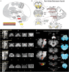
Brainstem. (A) Brain areas active during pain: periaqueductal gray (PAG), parabrachial nuclei (PB), rostral ventromedial medulla (RVM), spinal trigeminal nucleus (SpV) highlighted. (B) Schematic of brainstem nuclei associated with pain processing. Reprinted from PAIN Reports, Vol 4(4), V Napadow, R Sclocco, LA Henderson, Brainstem neuroimaging of nociception and pain circuitries, p e745, Copyright (2019) Napadow et al., under the Creative Commons Attribution License (CC-BY). DOI: https://dx.doi.org/10.1097%2FPR9.0000000000000745. (C) Axial slices containing brainstem nuclei from Figure. 12(B) arranged to compare the spatial resolution and quality of anatomical and functional MRI data at different magnetic field strengths (7 Tesla and 3 Tesla). Advances in imaging techniques and technologies promise to advance neuroimaging investigation of the brainstem as subtle differences in increasingly fine and detailed structures can be appreciated by MRI. Reprinted from PAIN Reports, Vol 4(4), V Napadow, R Sclocco, LA Henderson, Brainstem neuroimaging of nociception and pain circuitries, p e745, Copyright (2019) Napadow et al., under the Creative Commons Attribution License (CC-BY). DOI: https://dx.doi.org/10.1097%2FPR9.0000000000000745. (D) fMRI activations in the medulla, pons, and midbrain in response to brief noxious thermal stimulation, comprising activation of ascending nociceptive pathways and descending pain modulation, highlighting the dense and complex pain circuitry present in the brainstem. Myelin-stained ex-vivo axial sections are displayed to the right of corresponding sagittal and axial MRI slices. Reprinted from NeuroImage, Vol 124(Part A), AM Youssef, VG Macefield, LA Henderson, Pain inhibits pain; human brainstem mechanisms, p54-62, Copyright (2020), with permission from Elsevier. DOI: https://doi.org/10.1016/j.neuroimage.2015.08.060. DRN, dorsal raphe nucleus; DRt, dorsal reticular nucleus; LC, locus coeruleus; MRN, median raphe nucleus; NCF, nucleus cuneiformis; NGc, nucleus gigantocellularis; NRM, nucleus raphe magnus; NTS, nucleus tractus solitarii; PAG, periaqueductal gray; PBN, parabrachial nucleus; RVM, rostral ventromedial medulla; SpV, spinal trigeminal nucleus; VLM, ventrolateral medulla; SpVc, spinal trigeminal nucleus caudalis; SRD, subnucleus reticularis dorsalis; dlPons, dorsolateral pons; PAG, periaqueductal gray; SN, substantia nigra.
Figure 13
Figure 13
Thalamus. (A) Brain areas active during pain: Thalamus (Thal) highlighted. (B) An examination of resting state fMRI data finds that participants with orofacial pain, experimentally induced by orthodontic separators, have significantly different patterns and intensity of spontaneous neural activity (red=increased, blue=decreased) compared to controls; differences such as these support the notion that the thalamus has a greater role in somatosensory processing than simply relaying afferent signals. Reprinted from Frontiers in Neurology, Vol 11, Y Jin, H Yang, F Zhang, J Wang, H Liu, X Yang, H Long, F Li, Q Gong, W Lai, The Medial Thalamus Plays an Important Role in the Cognitive and Emotional Modulation of Orofacial Pain: A Functional Magnetic Resonance Imaging-Based Study, p589125, Copyright (2021) Jin et al., under the Creative Commons Attribution License (CC-BY). DOI: https://doi.org/10.3389/fneur.2020.589125. (C) The same orofacial pain participants from (B) were also found to have a widespread and significant reduction (yellow areas) in resting state functional connectivity between the medial thalamus and other brain areas, emphasizing the comprehensive brain-wide interconnections of the thalamus and its engagement in pain processing. Reprinted from Frontiers in Neurology, Vol 11, Y Jin, H Yang, F Zhang, J Wang, H Liu, X Yang, H Long, F Li, Q Gong, W Lai, The Medial Thalamus Plays an Important Role in the Cognitive and Emotional Modulation of Orofacial Pain: A Functional Magnetic Resonance Imaging-Based Study, p589125, Copyright (2021) Jin et al., under the Creative Commons Attribution License (CC-BY). DOI: https://doi.org/10.3389/fneur.2020.589125.
Figure 14
Figure 14
Consistent patterns of chronic pain. The ACC, PFC, and IC consistently display decreased grey matter in chronic pain conditions, along with impaired white matter health (FA) and opioid receptor binding. Chronic pain is also associated with reductions in N-acetyl aspartate in the ACC and PFC, while studies in rodents have found increased inflammation in these regions as well. Reprinted with permission from Springer Nature Customer Service Centre GmbH: Springer Nature, Nature Reviews Neuroscience, Vol 14, Cognitive and emotional control of pain and its disruption in chronic pain, MC Bushnell, M Ceko, LA Low, p502–11, Copyright (2013). DOI: https://doi.org/10.1038/nrn3516. Grey arrows, pain pathways; Black arrows, descending pathways; FA,functional anisotropy: a measure of molecule diffusion that serves as an index of white matter integrity; Opioids, opioid receptor binding: a marker of the ability to bind opioids and a way to analyze the health of descending pain systems; NAA, N-acetyl aspartate: a marker of neuronal viability. ACC, anterior cingulate cortex; PFC, prefrontal cortex; IC, insular cortex; S1, primary somatosensory cortex; S2, secondary somatosensory cortex; SPL, superior parietal lobe; BG, basal ganglia; AMY, Amygdala; PAG, periaqueductal gray; PB, parabrachial nuclei; RVM, rostral ventromedial medulla.

Similar articles

Cited by

References

    1. Raja SN, Carr DB, Cohen M, Finnerup NB, Flor H, Gibson S, et al. . The revised International Association for the Study of Pain definition of pain: concepts, challenges, and compromises. Pain. (2020) 161:1976–82. 10.1097/j.pain.0000000000001939 - DOI - PMC - PubMed
    1. Woolf CJ. What is this thing called pain? J Clin Invest. (2010) 120:3742–4. 10.1172/JCI45178 - DOI - PMC - PubMed
    1. Baliki MN, Apkarian AV. Nociception, pain, negative moods, and behavior selection. Neuron. (2015) 87:474–91. 10.1016/j.neuron.2015.06.005 - DOI - PMC - PubMed
    1. Melzack R, Casey KL. Sensory, motivational, and central control determinants of pain: a new conceptual model. The skin senses. (1968) 1:423–43.
    1. Valet M, Sprenger T, Boecker H, Willoch F, Rummeny E, Conrad B, et al. . Distraction modulates connectivity of the cingulo-frontal cortex and the midbrain during pain–an fMRI analysis. Pain. (2004) 109:399–408. 10.1016/j.pain.2004.02.033 - DOI - PubMed