

Neurothekeoma located in the hallux and axilla: Two case reports
- PMID: 35211617
- PMCID: PMC8855274
- DOI: 10.12998/wjcc.v10.i5.1738
Neurothekeoma located in the hallux and axilla: Two case reports
Abstract
Background: Neurothekeomas (NTKs) are rare benign soft tissue tumours that typically occur in the head, trunk, and upper limbs and are rare in other parts of the body.
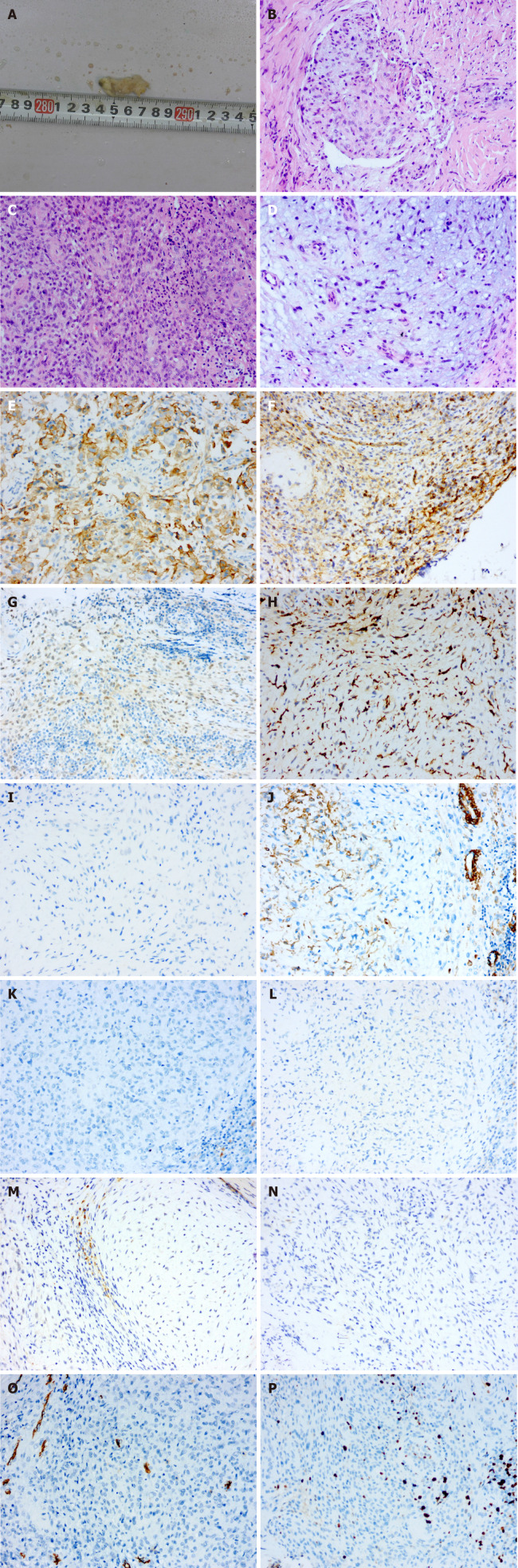
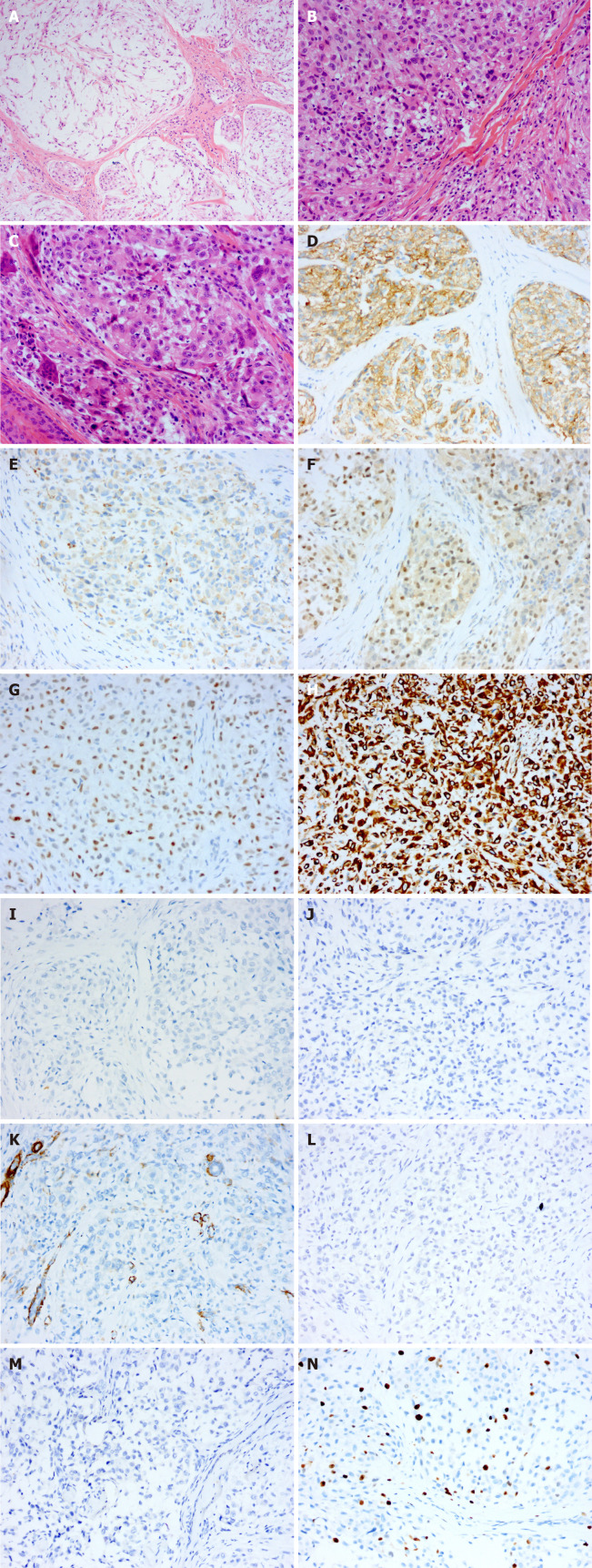
Case summary: Herein, we present two rare cases in which primary NTKs were located in the hallux and axilla. A 47-year-old woman complained of a verrucous bulge on the plantar side of the left hallux. The surface skin of the tumour was abraded due to poor wound healing. A 6-year-old boy complained of a gradually growing subcutaneous mass in the axilla. The tumours of both patients were completely resected, and the diagnosis of NTK was confirmed by histopathology. At the one-year follow-up, both patients had a good prognosis without local recurrence.
Conclusion: To date, NTKs located in the hallux and axilla have rarely been reported in the literature. We describe NTKs that occurred in unconventional areas and summarize the challenges in their diagnosis and differential diagnosis.
Keywords: Armpit; Case report; Hallux; Histopathological examination; Immunohistochemical staining; Neurothekeoma.
©The Author(s) 2022. Published by Baishideng Publishing Group Inc. All rights reserved.
Conflict of interest statement
Conflict-of-interest statement: The authors have no conflicts of interest to report.
Figures
Similar articles
-
Neurothekeoma of the Cornea.Ocul Oncol Pathol. 2016 Oct;2(4):212-217. doi: 10.1159/000444716. Epub 2016 Apr 1. Ocul Oncol Pathol. 2016. PMID: 27843897 Free PMC article.
-
Neurothekeoma of the eye, conjunctiva, and periorbital adnexa: A report of two cases and brief review.Surv Ophthalmol. 2019 Nov-Dec;64(6):852-857. doi: 10.1016/j.survophthal.2019.04.002. Epub 2019 Apr 9. Surv Ophthalmol. 2019. PMID: 30978337 Review.
-
[Clinicopathologic features of dermal nerve sheath myxoma and neurothekeoma: a comparative study].Zhonghua Bing Li Xue Za Zhi. 2016 Nov 8;45(11):755-761. doi: 10.3760/cma.j.issn.0529-5807.2016.11.003. Zhonghua Bing Li Xue Za Zhi. 2016. PMID: 27821229 Review. Chinese.
-
Unusual Recurrent Lateral Canthus Mass in a 16-Year-Old Male Patient: Neurothekeoma.Case Rep Oncol. 2019 Sep 17;12(3):693-697. doi: 10.1159/000502948. eCollection 2019 Sep-Dec. Case Rep Oncol. 2019. PMID: 31607885 Free PMC article.
-
Multiple cellular neurothekeomas in a middle-aged woman including the lower extremity: A case report and review of the current literature.J Cutan Pathol. 2019 Jan;46(1):67-73. doi: 10.1111/cup.13366. Epub 2018 Nov 8. J Cutan Pathol. 2019. PMID: 30270462 Review.
References
-
- Lau SK, Cassarino DS, Koh SS. Multiple myxoid cellular neurothekeomas in a patient with systemic lupus erythematosus. J Cutan Pathol . 2021;48:980–985. - PubMed
-
- Massimo JA, Gasibe M, Massimo I, Damilano CP, De Matteo E, Fiorentino J. Neurothekeoma: Report of two cases in children and review of the literature. Pediatr Dermatol . 2020;37:187–189. - PubMed
-
- Wiemeyer S, Hafer G. Neurothekeoma of the toe. Foot Ankle Spec . 2013;6:479–481. - PubMed
-
- Cavicchini S, Guanziroli E, Del Gobbo A, Scaparro M, Gianotti R. Neurothekeoma, a hard to diagnose neoplasm among red nodules. Australas J Dermatol . 2018;59:e280–e282. - PubMed
Publication types
LinkOut - more resources
Full Text Sources
