

Impact of Notch3 Activation on Aortic Aneurysm Development in Marfan Syndrome
- PMID: 35211631
- PMCID: PMC8863478
- DOI: 10.1155/2022/7538649
Impact of Notch3 Activation on Aortic Aneurysm Development in Marfan Syndrome
Abstract
Background: The leading cause of mortality in patients with Marfan syndrome (MFS) is thoracic aortic aneurysm and dissection. Notch signaling is essential for vessel morphogenesis and function. However, the role of Notch signaling in aortic pathology and aortic smooth muscle cell (SMC) differentiation in Marfan syndrome (MFS) is not completely understood.
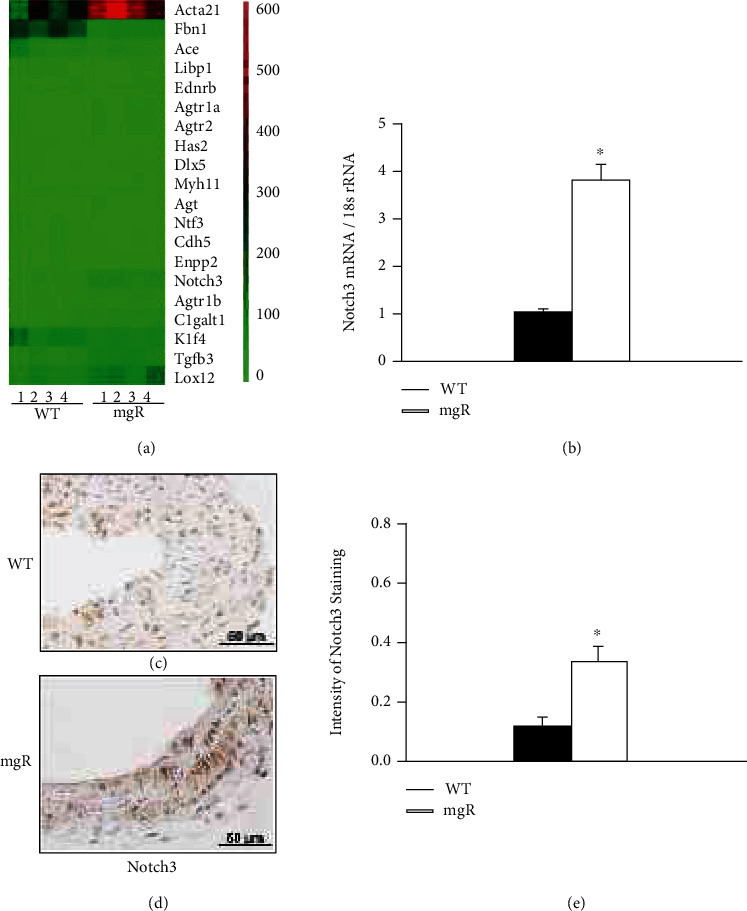
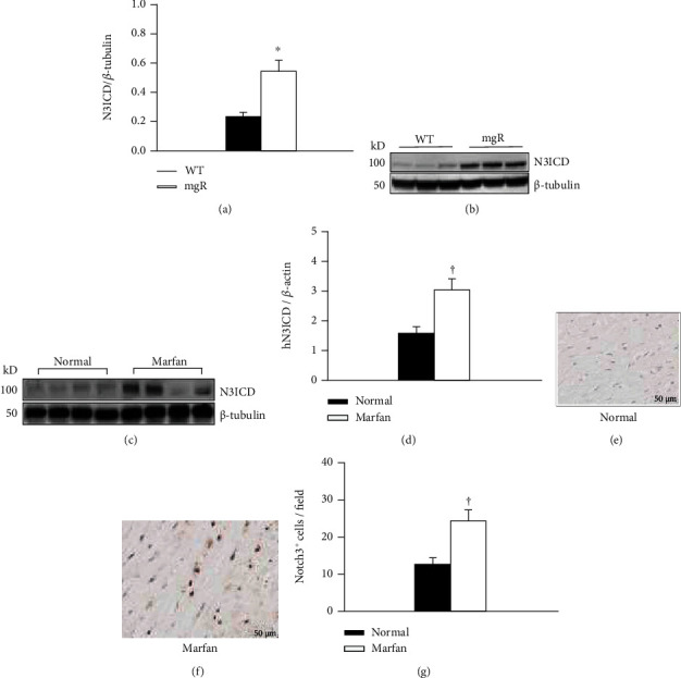
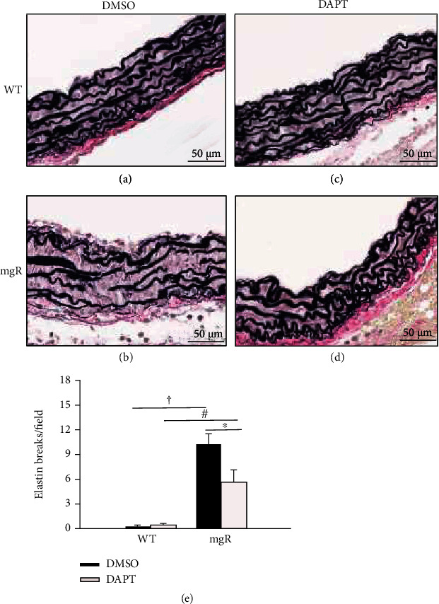
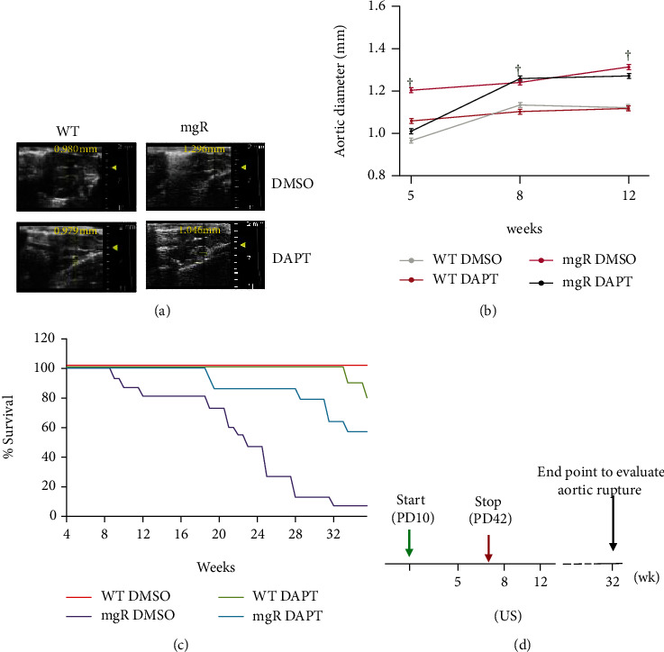
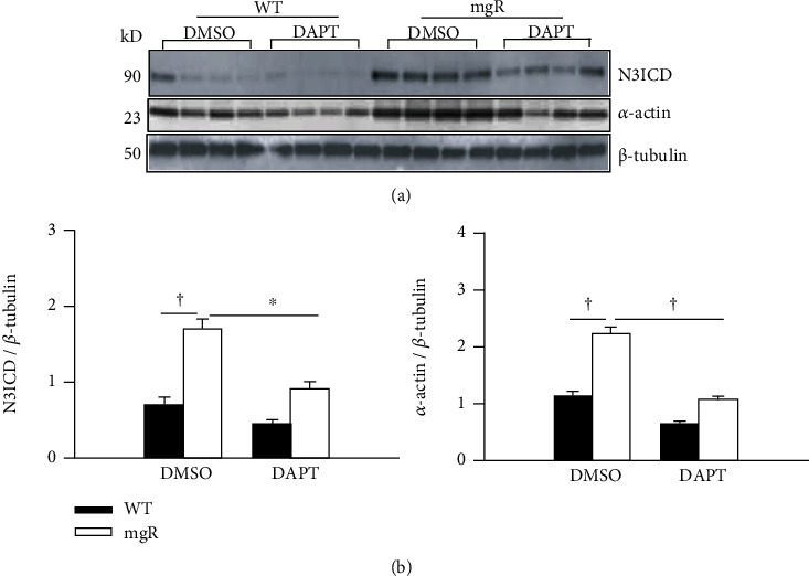
Methods: RNA-sequencing on ascending aortic tissue from a mouse model of MFS, Fbn1mgR/mgR , and wild-type controls was performed. Notch 3 expression and activation in aortic tissue were confirmed with real-time RT-PCR, immunohistochemistry, and Western blot. Fbn1mgR/mgR and wild-type mice were treated with a γ-secretase inhibitor, DAPT, to block Notch activation. Aortic aneurysms and rupture were evaluated with connective tissue staining, ultrasound, and life table analysis.
Results: The murine RNA-sequencing data were validated with mouse and human MFS aortic tissue, demonstrating elevated Notch3 activation in MFS. Data further revealed that upregulation and activation of Notch3 were concomitant with increased expression of SMC contractile markers. Inhibiting Notch3 activation with DAPT attenuated aortic enlargement and improved survival of Fbn1mgR/mgR mice. DAPT treatment reduced elastin fiber fragmentation in the aorta and reversed the differentiation of SMCs.
Conclusions: Our data demonstrated that matrix abnormalities in the aorta of MFS are associated with increased Notch3 activation. Enhanced Notch3 activation in MFS contributed to aortic aneurysm formation in MFS. This might be mediated by inducing a contractile phenotypic change of SMC. Our results suggest that inhibiting Notch3 activation may provide a strategy to prevent and treat aortic aneurysms in MFS.
Copyright © 2022 Kathryn Jespersen et al.
Conflict of interest statement
The authors declare that they have no conflicts of interest.
Figures

Similar articles
-
Single-Cell Transcriptomic Profiling of Vascular Smooth Muscle Cell Phenotype Modulation in Marfan Syndrome Aortic Aneurysm.Arterioscler Thromb Vasc Biol. 2020 Sep;40(9):2195-2211. doi: 10.1161/ATVBAHA.120.314670. Epub 2020 Jul 23. Arterioscler Thromb Vasc Biol. 2020. PMID: 32698686 Free PMC article.
-
Divergent effects of canonical and non-canonical TGF-β signalling on mixed contractile-synthetic smooth muscle cell phenotype in human Marfan syndrome aortic root aneurysms.J Cell Mol Med. 2020 Feb;24(3):2369-2383. doi: 10.1111/jcmm.14921. Epub 2019 Dec 30. J Cell Mol Med. 2020. PMID: 31886938 Free PMC article.
-
Resveratrol Inhibits Aortic Root Dilatation in the Fbn1C1039G/+ Marfan Mouse Model.Arterioscler Thromb Vasc Biol. 2016 Aug;36(8):1618-26. doi: 10.1161/ATVBAHA.116.307841. Epub 2016 Jun 9. Arterioscler Thromb Vasc Biol. 2016. PMID: 27283746 Free PMC article.
-
Insights into elastic fiber fragmentation: Mechanisms and treatment of aortic aneurysm in Marfan syndrome.Vascul Pharmacol. 2023 Dec;153:107215. doi: 10.1016/j.vph.2023.107215. Epub 2023 Aug 26. Vascul Pharmacol. 2023. PMID: 37640090 Free PMC article. Review.
-
Cardiovascular characteristics in Marfan syndrome and their relation to the genotype.Verh K Acad Geneeskd Belg. 2009;71(6):335-71. Verh K Acad Geneeskd Belg. 2009. PMID: 20232788 Review.
Cited by
-
Gamma Secretase Inhibitors as Potential Therapeutic Targets for Notch Signaling in Uterine Leiomyosarcoma.Int J Mol Sci. 2022 May 26;23(11):5980. doi: 10.3390/ijms23115980. Int J Mol Sci. 2022. PMID: 35682660 Free PMC article.
-
Molecular Mechanisms in Genetic Aortopathy-Signaling Pathways and Potential Interventions.Int J Mol Sci. 2023 Jan 16;24(2):1795. doi: 10.3390/ijms24021795. Int J Mol Sci. 2023. PMID: 36675309 Free PMC article. Review.
-
Contributions of Germline and Somatic Mosaic Genetics to Thoracic Aortic Aneurysms in Nonsyndromic Individuals.J Am Heart Assoc. 2024 Jul 16;13(14):e033232. doi: 10.1161/JAHA.123.033232. Epub 2024 Jul 3. J Am Heart Assoc. 2024. PMID: 38958128 Free PMC article.
-
Animal Models, Pathogenesis, and Potential Treatment of Thoracic Aortic Aneurysm.Int J Mol Sci. 2024 Jan 11;25(2):901. doi: 10.3390/ijms25020901. Int J Mol Sci. 2024. PMID: 38255976 Free PMC article. Review.
-
Marfan syndrome: insights from animal models.Front Genet. 2025 Jan 6;15:1463318. doi: 10.3389/fgene.2024.1463318. eCollection 2024. Front Genet. 2025. PMID: 39834548 Free PMC article. Review.
References
MeSH terms
Substances
LinkOut - more resources
Full Text Sources
Medical
Miscellaneous
