. 2022 Jan 19:24:268-279.
doi: 10.1016/j.omtm.2022.01.006. eCollection 2022 Mar 10.

Delivery of non-viral naked DNA vectors to liver in small weaned pigs by hydrodynamic retrograde intrabiliary injection

Affiliations

Delivery of non-viral naked DNA vectors to liver in small weaned pigs by hydrodynamic retrograde intrabiliary injection

Tatjana Chan et al. Mol Ther Methods Clin Dev. .

Abstract

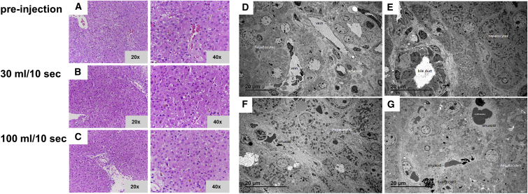
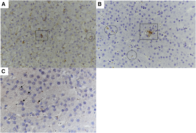
Hepatic gene therapy by delivering non-integrating therapeutic vectors in newborns remains challenging due to the risk of dilution and loss of efficacy in the growing liver. Previously we reported on hepatocyte transfection in piglets by intraportal injection of naked DNA vectors. Here, we established delivery of naked DNA vectors to target periportal hepatocytes in weaned pigs by hydrodynamic retrograde intrabiliary injection (HRII). The surgical procedure involved laparotomy and transient isolation of the liver. For vector delivery, a catheter was placed within the common bile duct by enterotomy. Under optimal conditions, no histological abnormalities were observed in liver tissue upon pressurized injections. The transfection of hepatocytes in all tested liver samples was observed with vectors expressing luciferase from a liver-specific promoter. However, vector copy number and luciferase expression were low compared to hydrodynamic intraportal injection. A 10-fold higher number of vector genomes and luciferase expression was observed in pigs using a non-integrating naked DNA vector with the potential for replication. In summary, the HRII application was less efficient (i.e., lower luciferase activity and vector copy numbers) than the intraportal delivery method but was significantly less distressful for the piglets and has the potential for injection (or re-injection) of vector DNA by endoscopic retrograde cholangiopancreatography.

Keywords: DNA-vector; bile duct; hydrodynamic intrabiliary injection; liver gene therapy; non-viral; pig.


Conflict of interest statement

The authors declare no competing interests.

Figures

None
Graphical abstract
Figure 1
Figure 1
Scheme of the common bile duct access for hydrodynamic retrograde intrabiliary infusion (HRII) and outflow examination via real-time X-ray (pig A1) (A) Scheme of a pig liver with vena portae, Arteriahepatica, vena cava caudalis, and the gallbladder, including biliary tract. Laparotomy and transient clamping (clamps) of vena portae, A. hepatica, vena cava caudalis, and the cystic duct (asterisk) was performed to target periportal hepatocytes. Access of the HRII procedure was performed by enterotomy and moving the catheter forward through the papilla duodeni major (not shown) into the common hepatic duct (black arrow in B). The position of the catheter was ensured and tightly fixed with clamps. (B) Pre-injection to check catheter position (the circle with a black arrow indicates the tip of the catheter). (C) Post-contrast medium injection while obstructing vena cava caudalis ( “clamping of one”) with no outflow visible (circle). (D) Post-injection of contrast medium (22 mL 0.9% NaCl plus 8 mL contrast medium) without clamping; outflow in direction of the heart visible (circle). (E) Post-injection of contrast medium with clamping of all 3 vessels with no outflow visible (circle).
Figure 2
Figure 2
Histological and TEM analyses of liver tissue before and after HRII (pig A2) (A–C) H&E staining of liver biopsies pre-injection (A) and after injection conditions with a sterile saline solution (i.e., 30 mL in 10 s) (B), followed by a second injection of 100 mL in 10 s (C). Mild hemosiderosis and mild cytoplasmatic vacuolation could be detected in biopsies after the consecutive pressurized injections with no other morphological damage. (D–G) Ultrastructure (TEM) analyses of pig liver upon consecutive injection of 2 different conditions with sterile saline solution. Injection conditions of either 30 mL or 100 mL in 10 s, each followed by clamping for 1 min, did not reveal any visible morphological abnormalities. Pre-injection (D), after injection of 30 mL in 10 s (E), and after injection of 100 mL in 10 s (F). Overview of a hepatic triad (arteriole, vein, and bile duct) after injection of 100 mL in 10 s (G). No alterations in ultrastructure were observed in any of the specimens analyzed. Magnification or scale bars are indicated in the figure.
Figure 3
Figure 3
Sketches of pig liver lobes and efficacy of MC.P3Luc3 naked DNA vector delivery and luciferase expression upon HRII Drawing of a caudal view of a pig liver with position of collected samples indicated by black dots (PCR+), white dots (PCR), and yellow circles (luciferase expression positive). Injection dosage in milligrams and time points of sacrifice in hours or days are indicated. For the various conditions, compare also to Table 1. (A) Pig B1 (176-37): Injection of 12 mg naked DNA vector resulted in 100% transfection rate (75 of 75 samples) and a luciferase expression in 45% of all of the tested samples (34 of 75). (B) Pig B3 (176-31): Injection of 12 mg naked DNA vector resulted in a 100% transfection rate (75 of 75 samples) and a luciferase expression of 1.33% (1 of 75 samples). (C) Pig B4 (176-26): Injection of 2 mg naked DNA vector resulted in an 85% transfection rate (64 of 75 samples) and 8% luciferase-positive samples (6 of 75). (D) Pig B5 (176-28): Injection of 2 mg naked DNA vector resulted in a transfection rate of 36% (27 of 75 samples), but undetectable luciferase expression. (E) Pig B6 (176-29): Injection of 12 mg naked DNA vector resulted in a 100% (74 of 74 samples) transfection rate and 9.5% luciferase expression (7 of 74 samples). (F) Pig B7 (176-30): Injection of 12 mg naked DNA vector showed a 100% (75 of 75 samples) transfection rate and a luciferase expression of 6.7% (5 of 75 samples). Note that “10 days” means between 9 and 11 days post-injection.
Figure 4
Figure 4
Sketches of pig liver lobes and efficacy of nSMARter.P3Luc1 naked DNA vector delivery and luciferase expression upon HRII The position of collected samples is indicated by dots; black dots are PCR+, white dots are PCR, and yellow circles are luciferase-positive expression. Injection dosage in milligrams and time points of sacrifice in hours or days are indicated. For the various conditions, compare also to Table 1. (A) Pig C2 (176-36): Injection of 12 mg naked DNA vector resulted in an equal luciferase expression of 85.3% (64 of 75 samples positive), with a flow rate of 100 mL in 10 s after 6 h. (B) Pig C3 (176-32): Injection of 12 mg naked DNA vector resulted in a luciferase expression of 4% (3 of 75 samples positive), with a flow rate of 100 mL in 10 s after 10 days. (C) Pig C4 (176-33): Injection of 12 mg naked DNA vector resulted in a luciferase expression of 2.67% (2 of 75 samples positive), with a reduced flow rate of 30 mL in 10 s after 10 days. A sample was positive above the threshold of 0.08 RLU/μg protein. Note that “10 days” means between 9 and 11 days post-injection.
Figure 5
Figure 5
Cryosections and formalin-fixed paraffin embedded pig liver for luciferase detection in hepatocytes (A and B) DAB-chromogen staining for luciferase with a positive reaction (matte brown signal) for hepatocytes (square). A positive reaction is also seen from hepatocellular bile or hemosiderin with crystal brown signals (circles). (C) AEC-chromogen is used to distinguish between hepatocellular bile and hemosiderin with a very faint red positive intracellular reaction (black arrows). AEC, 3-amino-9-ethylcarbazol chromogen; DAB, 3,3′-diaminobenzidine. Liver samples were from pig A2 (sacrificed on day 10 after vector injection).

Similar articles

Cited by

References

    1. Dunbar C.E., High K.A., Joung J.K., Kohn D.B., Ozawa K., Sadelain M. Gene therapy comes of age. Science. 2018;359:eaan4672. doi: 10.1126/science.aan4672. - DOI - PubMed
    1. Baruteau J., Waddington S.N., Alexander I.E., Gissen P. Gene therapy for monogenic liver diseases: clinical successes, current challenges and future prospects. J. Inherit. Metab. Dis. 2017;40:497–517. doi: 10.1007/s10545-017-0053-3. - DOI - PMC - PubMed
    1. Colella P., Ronzitti G., Mingozzi F. Emerging issues in AAV-mediated in vivo gene therapy. Mol. Ther. Methods Clin. Dev. 2018;8:87–104. doi: 10.1016/j.omtm.2017.11.007. - DOI - PMC - PubMed
    1. Mendell J.R. Current clinical applications of in vivo gene therapy with AAVs. Mol. Ther. 2021;29:464–488. doi: 10.1016/j.ymthe.2020.12.007. - DOI - PMC - PubMed
    1. Dave U.P., Cornetta K. AAV joins the rank of genotoxic vectors. Mol. Ther. 2021;29:418–419. doi: 10.1016/j.ymthe.2021.01.007. - DOI - PMC - PubMed

LinkOut - more resources