Review
. 2022 Apr;9(2):021907.
doi: 10.1117/1.NPh.9.2.021907. Epub 2022 Feb 18.

Photonics tools begin to clarify astrocyte calcium transients

Affiliations
Review

Photonics tools begin to clarify astrocyte calcium transients

Kelsea A Gorzo et al. Neurophotonics. 2022 Apr.

Abstract

Astrocytes integrate information from neurons and the microvasculature to coordinate brain activity and metabolism. Using a variety of calcium-dependent cellular mechanisms, these cells impact numerous aspects of neurophysiology in health and disease. Astrocyte calcium signaling is highly diverse, with complex spatiotemporal features. Here, we review astrocyte calcium dynamics and the optical imaging tools used to measure and analyze these events. We briefly cover historical calcium measurements, followed by our current understanding of how calcium transients relate to the structure of astrocytes. We then explore newer photonics tools including super-resolution techniques and genetically encoded calcium indicators targeted to specific cellular compartments and how these have been applied to astrocyte biology. Finally, we provide a brief overview of analysis software used to accurately quantify the data and ultimately aid in our interpretation of the various functions of astrocyte calcium transients.

Keywords: analysis; astrocyte; calcium; genetically encoded fluorescent calcium indicator; stimulation emission depletion; two-photon.


Figures

Fig. 1
Fig. 1
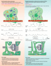
A comparison of small molecule calcium indicators with GECIs. A visual representation of the area of detected calcium signals inside the astrocyte is shown along with representative ΔF/F traces and general properties for various common indicators. A description of the advantages and disadvantages for small molecule indicators versus GECIs is also provided.
Fig. 2
Fig. 2
A schematic comparison of the neuro–glio–vascular interface as observed under diffraction-limited versus diffraction-limit exceeded microscopy for live-cell imaging. Diffraction limited tools (two-photon, visible confocal) are well suited to in vivo and brain slice preparations; however, only large astrocyte processes near synapses are resolved. Diffraction-limit exceeded microscopy (STED, 2P-STED, etc.) in acute slices or culture allow for specific compartments or organelles in addition to the loop-like/spongiform astrocyte arbor to be visualized.

References

    1. Araque A., et al. , “Gliotransmitters travel in time and space,” Neuron 81, 728–739 (2014).NERNET10.1016/j.neuron.2014.02.007 - DOI - PMC - PubMed
    1. Bezzi P., Volterra A., “A neuron-glia signalling network in the active brain,” Curr. Opin. Neurobiol. 11, 387–394 (2001).COPUEN10.1016/S0959-4388(00)00223-3 - DOI - PubMed
    1. Verkhratsky A., Nedergaard M., “Physiology of astroglia,” Physiol. Rev. 98, 239–389 (2018).PHREA710.1152/physrev.00042.2016 - DOI - PMC - PubMed
    1. Guerra-Gomes S., et al. , “Functional roles of astrocyte calcium elevations: from synapses to behavior,” Front. Cell. Neurosci. 11, 1–7 (2018).10.3389/fncel.2017.00427 - DOI - PMC - PubMed
    1. Volterra A., Liaudet N., Savtchouk I., “Astrocyte Ca2+ signalling: an unexpected complexity,” Nat. Rev. Neurosci. 15, 327–335 (2014).NRNAAN10.1038/nrn3725 - DOI - PubMed