. 2022 May;65(5):879-894.
doi: 10.1007/s00125-022-05650-4. Epub 2022 Feb 25.

Endothelial glycocalyx is damaged in diabetic cardiomyopathy: angiopoietin 1 restores glycocalyx and improves diastolic function in mice

Affiliations

Endothelial glycocalyx is damaged in diabetic cardiomyopathy: angiopoietin 1 restores glycocalyx and improves diastolic function in mice

Yan Qiu et al. Diabetologia. 2022 May.

Abstract

Aims/hypothesis: Diabetic cardiomyopathy (DCM) is a serious and under-recognised complication of diabetes. The first sign is diastolic dysfunction, which progresses to heart failure. The pathophysiology of DCM is incompletely understood but microcirculatory changes are important. Endothelial glycocalyx (eGlx) plays multiple vital roles in the microcirculation, including in the regulation of vascular permeability, and is compromised in diabetes but has not previously been studied in the coronary microcirculation in diabetes. We hypothesised that eGlx damage in the coronary microcirculation contributes to increased microvascular permeability and hence to cardiac dysfunction.

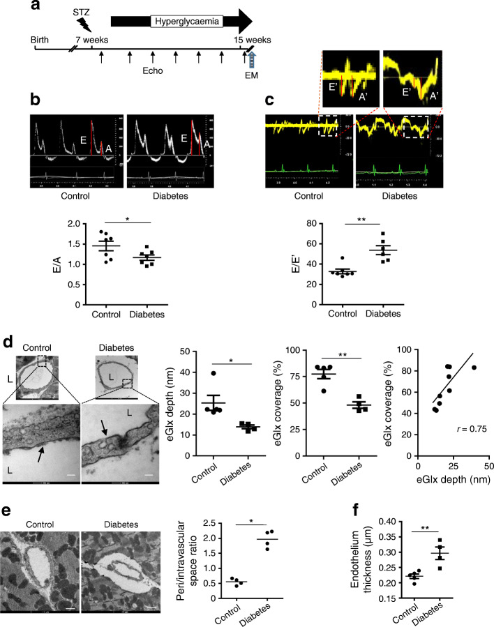
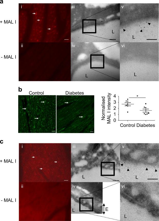
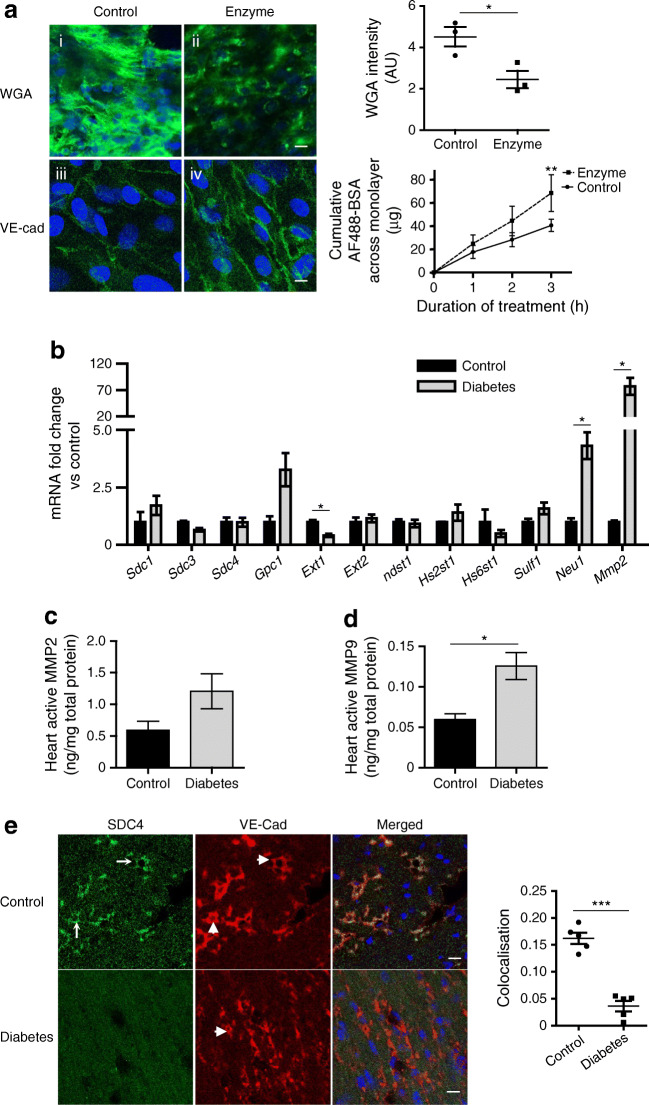
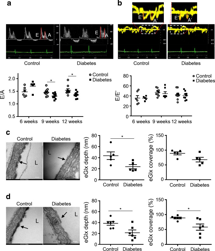
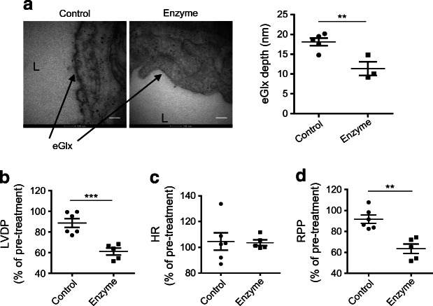
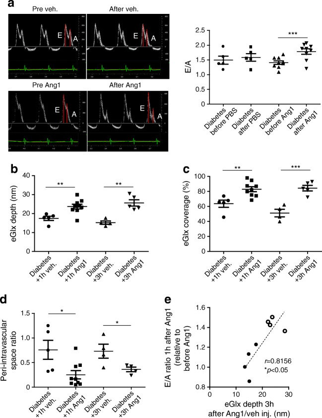
Methods: We investigated eGlx damage and cardiomyopathy in mouse models of type 1 (streptozotocin-induced) and type 2 (db/db) diabetes. Cardiac dysfunction was determined by echocardiography. We obtained eGlx depth and coverage by transmission electron microscopy (TEM) on mouse hearts perfusion-fixed with glutaraldehyde and Alcian Blue. Perivascular oedema was assessed from TEM images by measuring the perivascular space area. Lectin-based fluorescence was developed to study eGlx in paraformaldehyde-fixed mouse and human tissues. The eGlx of human conditionally immortalised coronary microvascular endothelial cells (CMVECs) in culture was removed with eGlx-degrading enzymes before measurement of protein passage across the cell monolayer. The mechanism of eGlx damage in the diabetic heart was investigated by quantitative reverse transcription-PCR array and matrix metalloproteinase (MMP) activity assay. To directly demonstrate that eGlx damage disturbs cardiac function, isolated rat hearts were treated with enzymes in a Langendorff preparation. Angiopoietin 1 (Ang1) is known to restore eGlx and so was used to investigate whether eGlx restoration reverses diastolic dysfunction in mice with type 1 diabetes.

Results: In a mouse model of type 1 diabetes, diastolic dysfunction (confirmed by echocardiography) was associated with loss of eGlx from CMVECs and the development of perivascular oedema, suggesting increased microvascular permeability. We confirmed in vitro that eGlx removal increases CMVEC monolayer permeability. We identified increased MMP activity as a potential mechanism of eGlx damage and we observed loss of syndecan 4 consistent with MMP activity. In a mouse model of type 2 diabetes we found a similar loss of eGlx preceding the development of diastolic dysfunction. We used isolated rat hearts to demonstrate that eGlx damage (induced by enzymes) is sufficient to disturb cardiac function. Ang1 restored eGlx and this was associated with reduced perivascular oedema and amelioration of the diastolic dysfunction seen in mice with type 1 diabetes.

Conclusions/interpretation: The association of CMVEC glycocalyx damage with diastolic dysfunction in two diabetes models suggests that it may play a pathophysiological role and the enzyme studies confirm that eGlx damage is sufficient to impair cardiac function. Ang1 rapidly restores the CMVEC glycocalyx and improves diastolic function. Our work identifies CMVEC glycocalyx damage as a potential contributor to the development of DCM and therefore as a therapeutic target.

Keywords: Angiopoietin 1; Coronary microcirculation; Diabetes; Glycocalyx; Permeability.


Figures

Fig. 1
Fig. 1
EGlx damage is associated with the development of DCM in FVB mice. (a) Diabetes was induced in FVB male mice by injection of low doses of STZ. The development of DCM was monitored with echocardiography for 9 weeks after STZ injection. (b, c) Representative pulsed wave Doppler images and reduced E/A ratio (b) and representative tissue Doppler images and increased E/E′ (c) in mice 9 weeks after STZ injection compared with control mice (n = 7 for control and 6 for diabetes; *p< 0.05 and **p< 0.01 [unpaired t test]). (d) Reduced eGlx depth and coverage in mice with DCM (n = 5; *p< 0.05 [unpaired t test]). Low- and high-magnification electron micrographs of capillaries from left ventricles of STZ-injected and control mice 9 weeks after STZ injection. Arrows show eGlx on top of endothelial cells. Scale bar, 100 nm. eGlx coverage (percentage of grid points at which eGlx depth is not less than 10 nm) is strongly positively associated with eGlx depth (n = 9; *p< 0.05 [Pearson r = 0.75]). (e, f) Increased peri/intravascular space ratio (area of perivascular space/area of intravascular space; n = 4; *p< 0.05 [unpaired t test]) (e) and endothelial thickness (n = 4 or 5; **p< 0.01 [unpaired t test]) (f) in the capillaries from diabetic vs non-diabetic hearts. Scale bar, 1 μm. Data are presented as mean ± SEM. EM, electron microscopy; L, capillary lumen
Fig. 2
Fig. 2
MAL I lectin staining confirms loss of eGlx in a mouse model of type 1 diabetes and demonstrates eGlx in human coronary capillaries. (a) MAL I lectin binds to sugar residues in the eGlx of FVB mouse coronary capillaries (i, ii show fluorescence microscopy images; iii–vi show TEM images). MAL I lectin-binding molecules mainly expressed in eGlx were confirmed by correlative light and electron microscopy using quantum dots binding to MAL I. Arrows point to coronary microvessels stained with MAL I. Arrowheads point to quantum dots associated with MAL I. (b) Reduced MAL I expression in left ventricles of mice with DCM (n = 7; *p< 0.05 [unpaired t test]). Arrows point to coronary microvessels stained with MAL I. (c) MAL I lectin binds to sugar residues in eGlx of human coronary capillaries (i, ii show fluorescence microscopy images; iii–vi show TEM images). Arrows point to coronary microvessels stained with MAL I. Arrowheads point to quantum dots associated with MAL I. (a, c) Scale bar, 15 μm in fluorescent images, 200 nm in electron microscopy images a(iii, iv) and c(iii, iv), and 100 nm in electron microscopy images a(v, vi) and c(v, vi). (b) Scale bars, 25 μm. Data are presented as mean ± SEM. E, endothelium; L, capillary lumen
Fig. 3
Fig. 3
Enzymatic eGlx disruption causes increased transendothelial permeability and myocardial MMP activity is increased in diabetes. The direct effect of eGlx damage on endothelial cell function was investigated on ciCMVECs. ciCMVECs were cultured until cells formed a confluent monolayer, then subjected to enzyme treatment (heparinase 1 U/ml + hyaluronidase 4.5 U/ml + chondroitinase 100 mU/ml) in serum-free medium for 3 h before BSA passage measurement. (a) Enzymatic treatment reduced eGlx, confirmed by reduced FITC–WGA binding (n = 3; *p< 0.05 [paired t test]), and enhanced BSA passage across the ciCMVEC monolayer (n = 3; **p< 0.01 [two-way ANOVA]) (i, ii). The ciCMVEC monolayer cell–cell junctions remained intact, as shown by maintained VE-cadherin junctional staining (iii, iv). Scale bar, 10 μm. (b) The mRNA expression levels of enzymes relevant to eGlx synthesis and shedding were investigated with TaqMan qRT-PCR array on endothelial cells collected by FACS from FVB mouse hearts 9 weeks after STZ injection. Mmp2 mRNA level was dramatically increased (n = 3; *p< 0.05 [unpaired t test]). (c, d) MMP2 (c) and MMP9 (d) activity in FVB mouse heart tissue homogenate were measured by activity assays. We found a significant increase in MMP9 activity in diabetic heart (n = 4; *p< 0.05 [unpaired t test]) and a non-significant increase in MMP2 activity (p = 0.11). (e) SDC4 was mainly expressed in coronary microvessels and its expression was reduced in left ventricles of mice with DCM (n = 5; ***p< 0.001 [unpaired t test]). Arrows point to SDC4 staining and arrowheads point to VE-cadherin staining. Scale bar, 10 μm. Data are presented as mean ± SEM. AF488, Alexa Fluor 488; AU, arbitrary units; VE-Cad, VE-cadherin
Fig. 4
Fig. 4
eGlx damage is associated with the development of DCM in a mouse model of type 2 diabetes. The development of DCM was monitored with echocardiography in db/db mice and control lean mice. (a) Representative pulsed wave Doppler images and reduced E/A ratio in diabetic mice when mice were 9 and 12 weeks old, compared with control lean mice (n = 6–9 for control mice and 5–9 for diabetic mice; *p< 0.05 [unpaired t test]). (b) Representative tissue Doppler images and no change of E/E′ in diabetic mice (n = 6–9 for control mice and 5–9 for diabetic mice). (c, d) At the age of 7 weeks (pre-DCM) (c) and 12 weeks (after the development of DCM) (d) control and diabetic mice were perfusion-fixed with Alcian Blue and glutaraldehyde solution for electron microscopy preparation. High-magnification electron micrographs of capillaries from left ventricles are presented. Scale bar, 100 nm. Arrows point to the eGlx on top of endothelial cells. Significantly reduced eGlx depth in heart capillaries was observed in 7-week-old diabetic mice (c; n = 5; *p< 0.05 [unpaired t test]). Significantly reduced eGlx depth and coverage (% of grid points with eGlx depth > 10 nm) were observed in heart capillaries from 12-week-old diabetic mice (d; n = 6; *p< 0.05 [unpaired t test]). Data are presented as mean ± SEM. L, capillary lumen
Fig. 5
Fig. 5
Damage of eGlx with enzyme treatment leads to impaired contractility in rat hearts. Hearts isolated from Sprague Dawley rats were perfused retrogradely with Krebs solution using Langendorff preparation. Once heart beats were stable, the hearts were further perfused with or without the combination of hyaluronidase (14 μg/ml) and chondroitinase (0.0022 U/ml) in Krebs solution for 40 min. (a) Damage to the eGlx after enzyme perfusion was identified in hearts perfusion-fixed with Alcian Blue and glutaraldehyde solution for electron microscopy preparation after heart functions were monitored. eGlx depth was decreased in the left ventricle capillaries of isolated rat hearts after enzyme perfusion (n = 5 for control and 3 for enzyme-perfused hearts; **p< 0.01 [unpaired t test]). Scale bar, 100 nm. (bd) Perfusion with the combination of enzymes caused reduced LVDP (***p< 0.001 [unpaired t test]) (b), left heart rate unchanged (c) and reduced rate pressure product (d), which is LVDP × HR (**p< 0.01 [unpaired t test]). n = 5 or 6. Data are presented as mean ± SEM. HR, heart rate; L, capillary lumen; LVDP, left ventricular developed pressure; RPP, rate pressure product
Fig. 6
Fig. 6
Ang1 improves diastolic heart function of diabetic FVB mice, associated with increased eGlx depth and coverage, and reduces perivascular space. Diabetes was induced in male FVB mice by STZ injections. Nine weeks after STZ injections, one group of diabetic mice were treated with Ang1 (200 ng/ml of blood volume, i.v.). Diastolic function was monitored before and 1 h after treatment. These mice were perfusion-fixed with Alcian Blue and glutaraldehyde solution for electron microscopy preparation 3 h after injection with Ang1. The effects of 1 h treatment with Ang1 or vehicle on eGlx were identified using another batch of mice. (a) One hour of Ang1 treatment improves diastolic heart function (n = 5 for diabetes+vehicle and 9 for diabetes+Ang1; ***p< 0.001 [unpaired t test]). (b, c) Electron microscopy shows increased eGlx depth at both 1 h (n = 5 for diabetes+vehicle and 9 for diabetes+Ang1; **p< 0.01 [unpaired t test]) and 3 h after Ang1 treatment (n = 4 for diabetes+vehicle and 5 for diabetes+Ang1; **p< 0.01 [unpaired t test]) (b), and increased eGlx coverage (**p< 0.01 and ***p< 0.001 [unpaired t test]) (c). (d) The enlarged perivascular space in mice with DCM is also reduced by Ang1 (n = 5 for diabetes+vehicle and 9 for diabetes+Ang1 for 1 h; n = 4 for diabetes+vehicle and diabetes+Ang1 for 3 h; *p< 0.05 [unpaired t test]). (e) The improvement of heart diastolic function in FVB mice by Ang1/vehicle is correlated with their corresponding eGlx depth identified 3 h after Ang1/vehicle treatments. Black circles, vehicle-treatment; white circles, Ang1 treatment (n = 8). Data are presented as mean ± SEM. Inj., injection; veh, vehicle

References

    1. Boyer JK, Thanigaraj S, Schechtman KB, Perez JE. Prevalence of ventricular diastolic dysfunction in asymptomatic, normotensive patients with diabetes mellitus. Am J Cardiol. 2004;93(7):870–875. doi: 10.1016/j.amjcard.2003.12.026. - DOI - PubMed
    1. Bell DS. Heart failure: the frequent, forgotten, and often fatal complication of diabetes. Diabetes Care. 2003;26(8):2433–2441. doi: 10.2337/diacare.26.8.2433. - DOI - PubMed
    1. Murtaza G, Virk HUH, Khalid M, et al. Diabetic cardiomyopathy - a comprehensive updated review. Prog Cardiovasc Dis. 2019;62(4):315–326. doi: 10.1016/j.pcad.2019.03.003. - DOI - PubMed
    1. Liu JE, Robbins DC, Palmieri V, et al. Association of albuminuria with systolic and diastolic left ventricular dysfunction in type 2 diabetes: the strong heart study. J Am Coll Cardiol. 2003;41(11):2022–2028. doi: 10.1016/S0735-1097(03)00403-0. - DOI - PubMed
    1. Arnold JM, Yusuf S, Young J, et al. Prevention of heart failure in patients in the heart outcomes prevention evaluation (HOPE) study. Circulation. 2003;107(9):1284–1290. doi: 10.1161/01.CIR.0000054165.93055.42. - DOI - PubMed

Publication types