. 2022 Mar 21;50(5):2959-2972.
doi: 10.1093/nar/gkac091.

The first crystal structures of hybrid and parallel four-tetrad intramolecular G-quadruplexes

Affiliations

The first crystal structures of hybrid and parallel four-tetrad intramolecular G-quadruplexes

Dana Beseiso et al. Nucleic Acids Res. .

Abstract

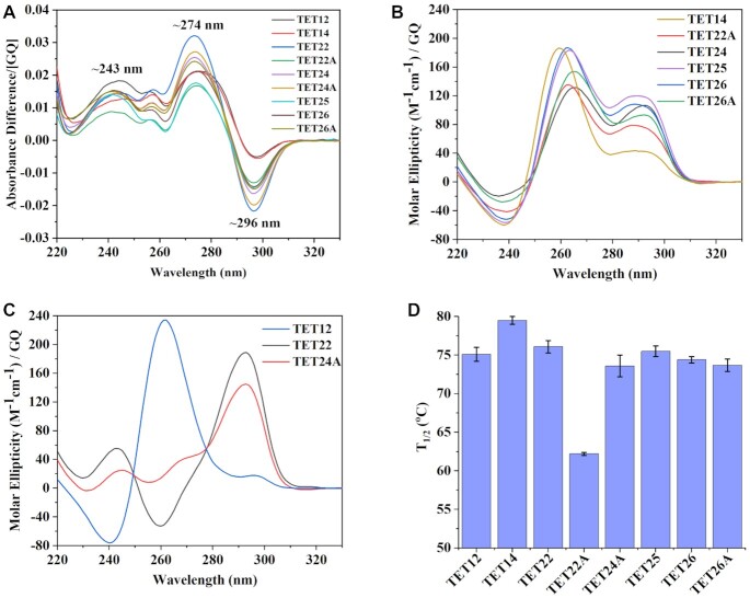
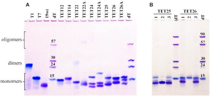
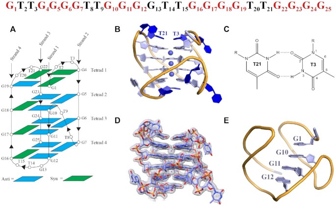
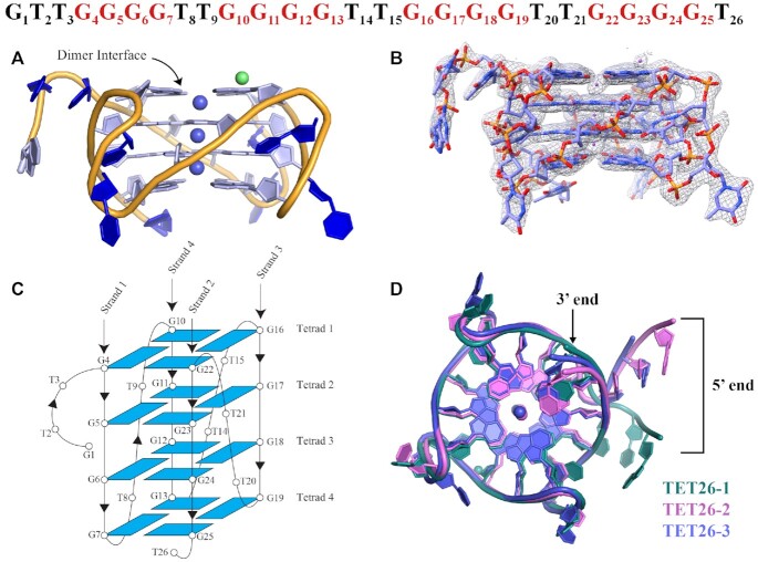
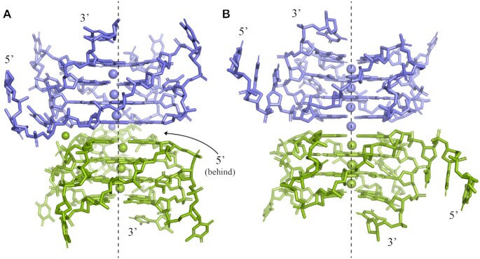
G-quadruplexes (GQs) are non-canonical DNA structures composed of stacks of stabilized G-tetrads. GQs play an important role in a variety of biological processes and may form at telomeres and oncogene promoters among other genomic locations. Here, we investigate nine variants of telomeric DNA from Tetrahymena thermophila with the repeat (TTGGGG)n. Biophysical data indicate that the sequences fold into stable four-tetrad GQs which adopt multiple conformations according to native PAGE. Excitingly, we solved the crystal structure of two variants, TET25 and TET26. The two variants differ by the presence of a 3'-T yet adopt different GQ conformations. TET25 forms a hybrid [3 + 1] GQ and exhibits a rare 5'-top snapback feature. Consequently, TET25 contains four loops: three lateral (TT, TT, and GTT) and one propeller (TT). TET26 folds into a parallel GQ with three TT propeller loops. To the best of our knowledge, TET25 and TET26 are the first reported hybrid and parallel four-tetrad unimolecular GQ structures. The results presented here expand the repertoire of available GQ structures and provide insight into the intricacy and plasticity of the 3D architecture adopted by telomeric repeats from T. thermophila and GQs in general.


Figures

Figure 1.
Figure 1.
Biophysical characterization of TET sequences. (A) TDS. (BC) CD scans at 20°C. (D) Thermal stability determined via CD melts. All samples were prepared in a 10K buffer at ∼4 μM per GQ.
Figure 2.
Figure 2.
Homogeneity, molecularity, and conformation of TET GQs via native PAGE. Fifteen percent gel was prepared in 1 × TBE supplemented with 10 mM KCl. (A) The DNA samples were prepared in 10K buffer at ∼0.10–0.24 mM. Size markers correspond to dTn sequences. Controls include T1, T7, and 19wt. (B) The DNA samples were prepared the following way: lane 1 – 0.10 mM DNA in 10K buffer; lane 2 – crystals dissolved in 10K buffer; and lane 3 – concentrated DNA sample used to grow crystals deposited in lane 2.
Figure 3.
Figure 3.
Crystal structure of the four-tetrad [3 + 1] hybrid TET25 GQ. The DNA sequence with nucleotide numbers is shown at the top. Guanines that participate in G-tetrad formation are colored in red. (A) Schematic representation of the folding topology with numbering schemes for nucleotides. Blue and green rectangles indicate anti and syn conformations of guanine bases, respectively. Chain orientation is indicated by arrowheads. (B) Cartoon representation of chain A with purines, pyrimidines, and sugars shown as filled rings and K+ ions as spheres. (C) Non-canonical T3-T21 base pair that stabilizes the GQ and maintains the position of the top lateral loops. (D) Chain A surrounded by the electron density at I/σ = 1.0. (E) 5′-top snapback.
Figure 4.
Figure 4.
Crystal structure of TET26. (A) Cartoon representation of the crystal structure of TET26-1 with nucleotides and sugars shown as filled rings. Purple spheres represent K+ ions and a green sphere represents a Na+ ion. (B) TET26-1 surrounded by the electron density at I/σ = 1.0. (C) Schematic representation of the TET26 folding topology with numbering schemes for the nucleotides. All nucleotides adopt anti glycosidic conformation. Chain orientation is indicated by arrowheads. (D) Comparison of TET26-1 (green), TET26-2 (pink) and TET26-3 (purple). An overlay of the three GQ structures shows that the GQ cores and loops are nearly identical whereas the 5′-GTT and 3′-T overhangs exhibit noticeable differences.
Figure 5.
Figure 5.
A depiction of (A) TET26-1 and (B) TET26-2 dimers with one monomer colored in purple and another in green. The symmetry related GQ was generated in PyMol. Two separate K+ channels exist in TET26-1. TET26-2 has one ion channel that houses seven K+ ions, including the K+ at the dimer interface which is modeled at 0.5 occupancy and colored in purple. The arrangement of TET26-3 is similar to that of TET26-2. The dashed line is drawn to guide the eye.

References

    1. Bhattacharyya D., Mirihana Arachchilage G., Basu S.. Metal cations in G-quadruplex folding and stability. Front. Chem. 2016; 4:38. - PMC - PubMed
    1. Mukundan V.T., Phan A.T.. Bulges in G-quadruplexes: broadening the definition of G-quadruplex-forming sequences. J. Am. Chem. Soc. 2013; 135:5017–5028. - PubMed
    1. Collie G.W., Campbell N.H., Neidle S.. Loop flexibility in human telomeric quadruplex small-molecule complexes. Nucleic Acids Res. 2015; 43:4785–4799. - PMC - PubMed
    1. Guédin A., Gros J., Alberti P., Mergny J.-L.. How long is too long? Effects of loop size on G-quadruplex stability. Nucleic Acids Res. 2010; 38:7858–7868. - PMC - PubMed
    1. Hazel P., Huppert J., Balasubramanian S., Neidle S.. Loop-length-dependent folding of G-quadruplexes. J. Am. Chem. Soc. 2004; 126:16405–16415. - PubMed

Publication types