How to stain nucleic acids and proteins in Miller spreads
- PMID: 35212500
- PMCID: PMC8883610
- DOI: 10.4081/ejh.2022.3364
How to stain nucleic acids and proteins in Miller spreads
Abstract
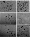
The spread technique proposed by Miller and Beatty in 1969 allowed for the first time the visualization at transmission electron microscopy of nucleic acids and chromatin in an isolated and distended conformation. The final step of staining the spread chromatin is of critical importance because it can strongly influence the interpretation of the results. We evaluated different staining techniques and the most part of them provided a good result. Specifically, well contrasted micrographs were obtained when staining with H3PW12O40 (PTA), as originally proposed by Miller and Beatty, and with two alternatives proposed here: uranyl acetate or terbium citrate staining. Quite good contrast of the spread DNA could be achieved also by using Osmium Ammine; while no or few contrast of nucleic acids was observed by staining with KMnO₄ and H3PMo12O40 (PMA) respectively.
Figures


Similar articles
-
The use of bismuth as an electron stain for nucleic acids.J Cell Biol. 1963 Apr;17(1):93-103. doi: 10.1083/jcb.17.1.93. J Cell Biol. 1963. PMID: 14011708 Free PMC article.
-
INTRAMITOCHONDRIAL FIBERS WITH DNA CHARACTERISTICS. I. FIXATION AND ELECTRON STAINING REACTIONS.J Cell Biol. 1963 Dec;19(3):593-611. doi: 10.1083/jcb.19.3.593. J Cell Biol. 1963. PMID: 14086138 Free PMC article.
-
Preferential staining of nucleic acid-containing structures for electron microscopy.J Biophys Biochem Cytol. 1961 Nov;11(2):273-96. doi: 10.1083/jcb.11.2.273. J Biophys Biochem Cytol. 1961. PMID: 14450292 Free PMC article.
-
Alternative staining methods for Lowicryl sections.J Histochem Cytochem. 1992 Jan;40(1):123-33. doi: 10.1177/40.1.1370308. J Histochem Cytochem. 1992. PMID: 1370308
-
Methods in ultrastructural cytochemistry of the cell nucleus.Prog Histochem Cytochem. 1980;13(1):1-72. doi: 10.1016/s0079-6336(80)80008-8. Prog Histochem Cytochem. 1980. PMID: 6153811 Review.
References
-
- Walter J, Joffe B, Bolzer A, Albiez H, Benedetti PA, Müller S., et al. . Towards many colors in FISH on 3D-preserved interphase nuclei. Cytogenet Genome Res 2006;114:367-78. - PubMed
-
- Lakadamyali M, Cosma MP. Advanced microscopy methods for visualizing chromatin structure. FEBS Lett 2015;589:3023-30. - PubMed
-
- Miller OL, Beatty BR. Visualization of nucleolar genes. Science 1969;164:955-7. - PubMed
MeSH terms
Substances
LinkOut - more resources
Full Text Sources

