. 2022 Mar 7;219(3):e20211275.
doi: 10.1084/jem.20211275. Epub 2022 Feb 25.

Glymphatic system clears extracellular tau and protects from tau aggregation and neurodegeneration

Affiliations

Glymphatic system clears extracellular tau and protects from tau aggregation and neurodegeneration

Kazuhisa Ishida et al. J Exp Med. .

Abstract

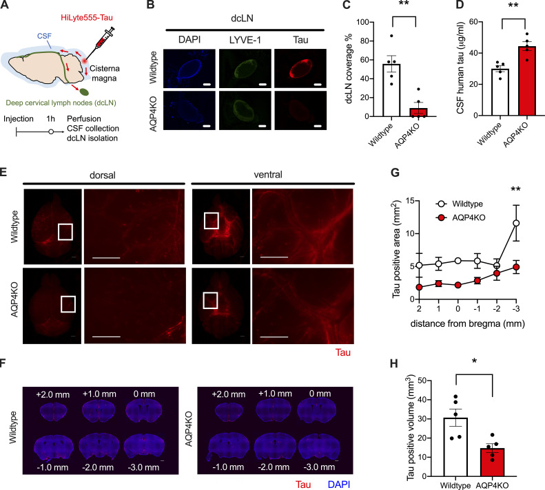
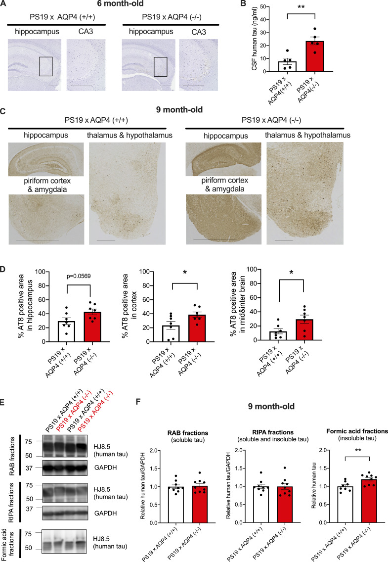
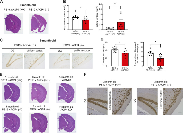
Accumulation of tau has been implicated in various neurodegenerative diseases termed tauopathies. Tau is a microtubule-associated protein but is also actively released into the extracellular fluids including brain interstitial fluid and cerebrospinal fluid (CSF). However, it remains elusive whether clearance of extracellular tau impacts tau-associated neurodegeneration. Here, we show that aquaporin-4 (AQP4), a major driver of the glymphatic clearance system, facilitates the elimination of extracellular tau from the brain to CSF and subsequently to deep cervical lymph nodes. Strikingly, deletion of AQP4 not only elevated tau in CSF but also markedly exacerbated phosphorylated tau deposition and the associated neurodegeneration in the brains of transgenic mice expressing P301S mutant tau. The current study identified the clearance pathway of extracellular tau in the central nervous system, suggesting that glymphatic clearance of extracellular tau is a novel regulatory mechanism whose impairment contributes to tau aggregation and neurodegeneration.


Conflict of interest statement

Disclosures: K. Yamada reported “part of the study was supported by a collaborative grant from NIPRO Co.” No other disclosures were reported.

Figures

None
Graphical abstract
Figure 1.
Figure 1.
Tau is cleared from the brain to CSF in an AQP4-dependent manner. (A) HiLyte 555–labeled human tau was stereotaxically injected into the brain. At 6, 12, 24, and 48 h after injection, the remaining tau in the brain and CSF tau levels were assessed. (B) Representative brain sections around the injection site with DAPI counterstaining. Scale bars, 1 mm. (C) Tau-positive area (mm2) was plotted with arrows that indicate injection sites. WT mice (n = 5), AQP4 KO mice (n = 6 for 12 h and 24 h, n = 5 for 6 h and 48 h). (D) Tau-positive volume (mm3) at different time points from injection. Two-way ANOVA with Bonferroni post-hoc analysis; *, P < 0.05; **, P < 0.01. n = 5/group. (E) CSF human tau levels at 6 (n = 5), 12 (n = 5), 24 (n = 5), and 48 h (n = 6) with hypothetical human tau concentration at 0 h were plotted. Two-way ANOVA with Bonferroni post-hoc analysis; ***, P < 0.001.
Figure 2.
Figure 2.
Tau clearance from CSF to dcLNs and influx into the brain occur in an AQP4-dependent manner. (A) At 1 h from intracisternal injection of HiLyte 555–labeled tau, CSF, brains, and dcLNs were analyzed. (B) Representative images of tau accumulation (red) in LYVE-1–positive dcLNs (pseudocolored green) with DAPI counterstaining (blue). Scale bars, 1 mm. (C) Percent area covered by tau in dcLNs. Unpaired two-tailed test; **, P < 0.01. n = 5/group. (D) CSF tau levels. Unpaired two-tailed test; **, P < 0.01. n = 5/group. (E) Representative images of tau accumulation (red). Scale bars, 1 mm. (F) Representative brain sections showing tau (red) accumulation with DAPI staining (blue). Scale bars, 1 mm. (G) Tau-positive area (mm2). Two-way ANOVA with Bonferroni post-hoc analysis; **, P < 0.01. n = 5/group. (H) Average tau-positive volume (mm3). Unpaired two-tailed test; *, P < 0.05. n = 5/group.
Figure S1.
Figure S1.
Ovalbumin influx from CSF to brain in WT mice and AQP4 KO mice. (A) Representative dorsal and ventral images of ovalbumin (green) and dextran (red) accumulation in WT mice or AQP4 KO mice. Scale bars, 1 mm. (B) Ovalbumin-positive area (mm2) was plotted from different distances to the bregma of WT mice and AQP4 KO mice. WT mice (n = 3), AQP4 KO mice (n = 4). Two-way ANOVA with Bonferroni post-hoc analysis; **, P < 0.01; ***, P < 0.001. (C) Ovalbumin-positive brain volume (mm3) at 1 h from injection in WT mice and AQP4 KO mice. Unpaired two-tailed t test; **, P < 0.01. WT mice (n = 3), AQP4 KO mice (n = 4).
Figure 3.
Figure 3.
AQP4 deficiency markedly exacerbates tau pathology in PS19 mice. (A) Representative images of AT8 staining in 6-month-old mice with hematoxylin staining. Scale bars, 300 μm. (B) CSF human tau levels in 6-month-old mice. Unpaired two-tailed t test; **, P < 0.01. n = 5/group. (C) Representative images of AT8 staining in 9-month-old mice. Scale bars, 1 mm for the hippocampus, piriform cortex, and amygdala and 300 μm for the thalamus and hypothalamus. (D) Quantification of the percentage of area covered by AT8 staining in 9-month-old mice. Unpaired two-tailed t test; *, P < 0.05. n = 7/group. (E) Representative immunoblots probing for human tau and GAPDH in RAB, RIPA, and formic acid fractions of 9-month-old PS19 × AQP4 (+/+) and PS19 × AQP4 (−/−) mice (in kD). (F) Quantification of immunoblot probing for human tau in RAB, RIPA, and formic acid fractions of 9-month-old PS19 × AQP4 (+/+) (n = 8 or 9) and PS19 × AQP4 (−/−) mice (n = 9). Unpaired two-tailed t test; **, P < 0.01. Source data are available for this figure: SourceData F3.
Figure S2.
Figure S2.
AQP4 deficiency markedly increases tau pathology stained with PHF-1 and MC1 in PS19 mice. (A) Representative images of 9-month-old PS19 × AQP4 (+/+), PS19 × AQP4 (+/−), and PS19 × AQP4 (−/−) mice stained with PHF-1. Scale bars, 1 mm. (B) Representative images of 9-month-old PS19 × AQP4 (+/+) and PS19 × AQP4 (−/−) mice stained with MC1. Scale bars, 300 μm. (C) Quantification of the percentage of area covered by MC1 staining in 9-month-old mice. Unpaired two-tailed t test; *, P < 0.05. n = 7/group.
Figure S3.
Figure S3.
LRP1 expression was comparable in PS19 mice regardless of AQP4 genotypes. (A) Representative images of 9-month-old PS19 × AQP4 (+/+) and PS19 × AQP4 (−/−) mice stained with LRP1 or AQP4. Arrows indicate neuronal staining and arrowheads indicate vasculatures. Scale bars, 20 μm. (B and C) Representative immunoblots (in kD) probing for LRP1 and actin in RIPA fractions of 9-month-old PS19 × AQP4 (+/+) (n = 9) and PS19 × AQP4 (−/−) (n = 9) and its quantification. Unpaired two-tailed t test; n.s., not significant. Source data are available for this figure: SourceData FS3.
Figure 4.
Figure 4.
AQP4 deficiency markedly exacerbates neurodegeneration in PS19 mice. (A) Representative images of 9-month-old mice stained with H&E staining. Scale bars, 1 mm. (B) Quantification of volume of hippocampus and lateral ventricle in 9-month-old mice. Unpaired two-tailed test; *, P < 0.05. n = 7/group. (C) Representative images of NeuN staining in DG and piriform cortex of 9-month-old mice. Scale bars, 100 μm. (D) Quantification of the thickness of granule cell layer in DG and the pyramidal cell layer in the piriform cortex of 9-month-old mice. Unpaired two-tailed test; *, P < 0.05. n = 7/group. (E) Representative images of 3- and 6-month-old PS19 × AQP4 (+/+) mice and 14-month-old WT mice or AQP4 KO mice stained with H&E. Scale bars, 1 mm. (F) Representative images of NeuN staining in 3-month-old PS19 × AQP4 (+/+) and PS19 × AQP4 (−/−) mice counter-stained with hematoxylin. Scale bars, 100 μm.

References

    1. Abe, Y., Ikegawa N., Yoshida K., Muramatsu K., Hattori S., Kawai K., Murakami M., Tanaka T., Goda W., Goto M., et al. . 2020. Behavioral and electrophysiological evidence for a neuroprotective role of aquaporin-4 in the 5xFAD transgenic mice model. Acta Neuropathol. Commun. 8:67. 10.1186/s40478-020-00936-3 - DOI - PMC - PubMed
    1. Aoyagi, H., Hasegawa M., and Tamaoka A.. 2007. Fibrillogenic nuclei composed of P301L mutant tau induce elongation of P301L tau but not wild-type tau. J. Biol. Chem. 282:20309–20318. 10.1074/jbc.M611876200 - DOI - PubMed
    1. Aspelund, A., Antila S., Proulx S.T., Karlsen T.V., Karaman S., Detmar M., Wiig H., and Alitalo K.. 2015. A dural lymphatic vascular system that drains brain interstitial fluid and macromolecules. J. Exp. Med. 212:991–999. 10.1084/jem.20142290 - DOI - PMC - PubMed
    1. Barthélemy, N.R., Horie K., Sato C., and Bateman R.J.. 2020a. Blood plasma phosphorylated-tau isoforms track CNS change in Alzheimer’s disease. J. Exp. Med. 217:1–12:e20200861. 10.1084/JEM.20200861 - DOI - PMC - PubMed
    1. Barthélemy, N.R., Li Y., Joseph-Mathurin N., Gordon B.A., Hassenstab J., Benzinger T.L.S., Buckles V., Fagan A.M., Perrin R.J., Goate A.M., et al. 2020b. A soluble phosphorylated tau signature links tau, amyloid and the evolution of stages of dominantly inherited Alzheimer’s disease. Nat. Med. 26:398–407. 10.1038/s41591-020-0781-z - DOI - PMC - PubMed

Publication types

MeSH terms