. 2022 Nov 10;118(14):2960-2972.
doi: 10.1093/cvr/cvac023.

Mapping the developing human cardiac endothelium at single-cell resolution identifies MECOM as a regulator of arteriovenous gene expression

Affiliations

Mapping the developing human cardiac endothelium at single-cell resolution identifies MECOM as a regulator of arteriovenous gene expression

Ian R McCracken et al. Cardiovasc Res. .

Abstract

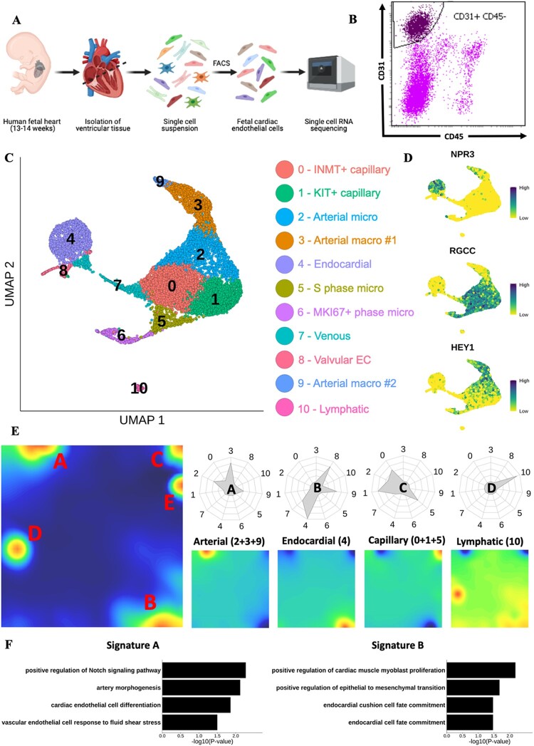
Aims: Coronary vasculature formation is a critical event during cardiac development, essential for heart function throughout perinatal and adult life. However, current understanding of coronary vascular development has largely been derived from transgenic mouse models. The aim of this study was to characterize the transcriptome of the human foetal cardiac endothelium using single-cell RNA sequencing (scRNA-seq) to provide critical new insights into the cellular heterogeneity and transcriptional dynamics that underpin endothelial specification within the vasculature of the developing heart.

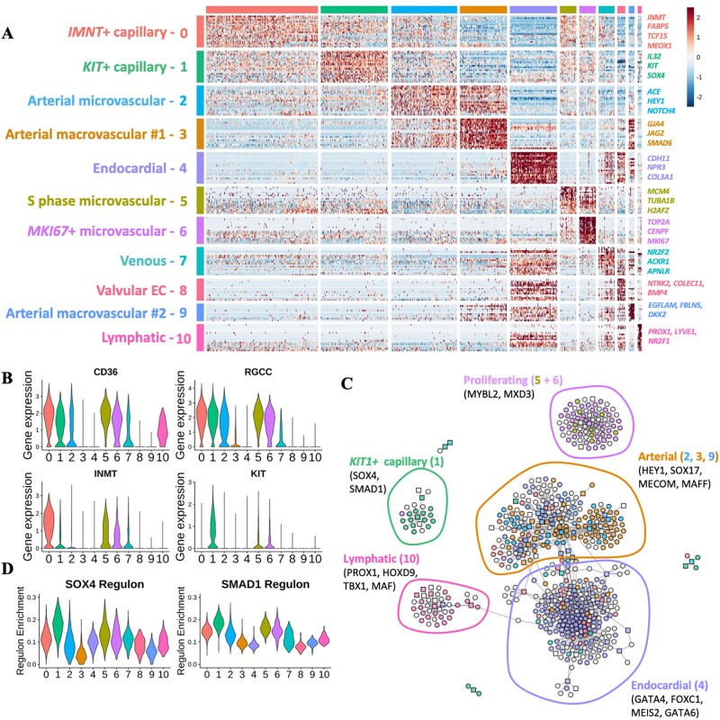
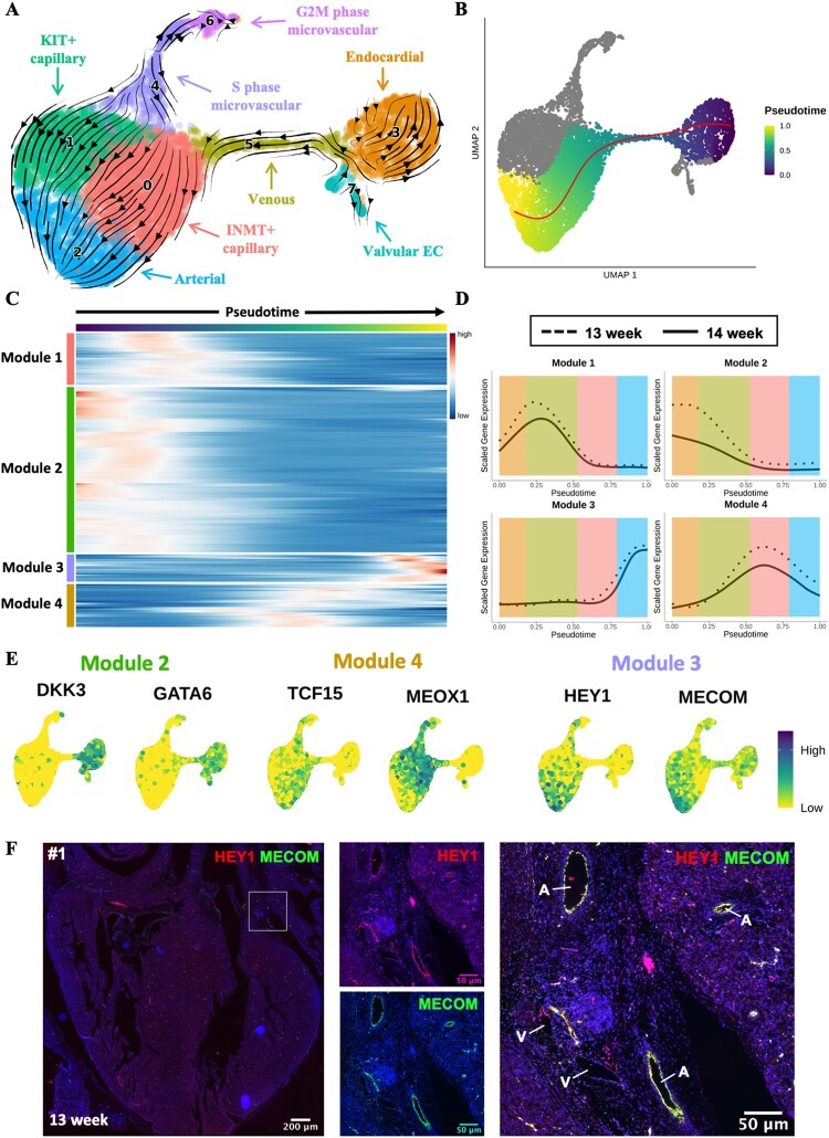
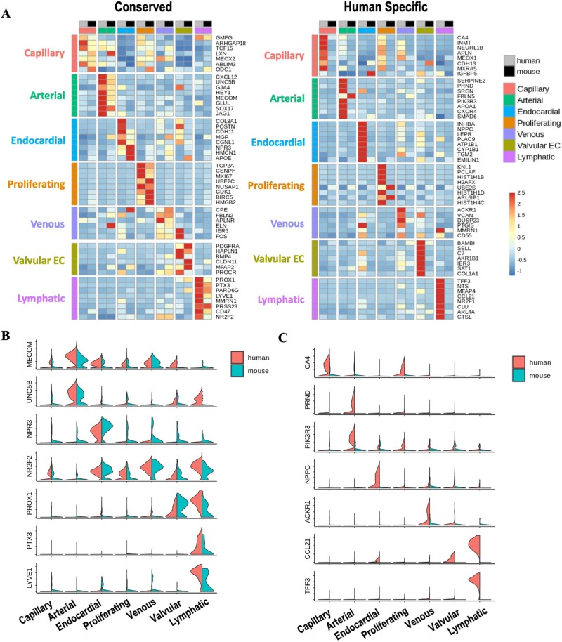
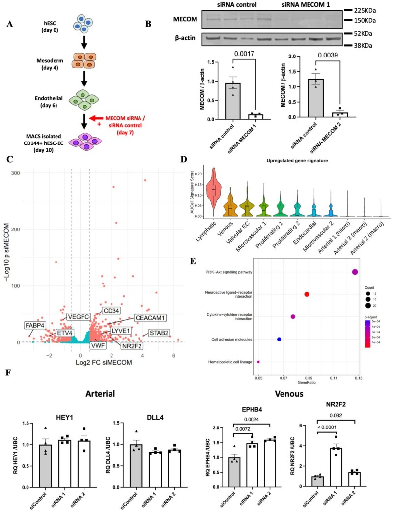
Methods and results: We acquired scRNA-seq data of over 10 000 foetal cardiac endothelial cells (ECs), revealing divergent EC subtypes including endocardial, capillary, venous, arterial, and lymphatic populations. Gene regulatory network analyses predicted roles for SMAD1 and MECOM in determining the identity of capillary and arterial populations, respectively. Trajectory inference analysis suggested an endocardial contribution to the coronary vasculature and subsequent arterialization of capillary endothelium accompanied by increasing MECOM expression. Comparative analysis of equivalent data from murine cardiac development demonstrated that transcriptional signatures defining endothelial subpopulations are largely conserved between human and mouse. Comprehensive characterization of the transcriptional response to MECOM knockdown in human embryonic stem cell-derived EC (hESC-EC) demonstrated an increase in the expression of non-arterial markers, including those enriched in venous EC.

Conclusions: scRNA-seq of the human foetal cardiac endothelium identified distinct EC populations. A predicted endocardial contribution to the developing coronary vasculature was identified, as well as subsequent arterial specification of capillary EC. Loss of MECOM in hESC-EC increased expression of non-arterial markers, suggesting a role in maintaining arterial EC identity.

Keywords: Coronary vasculature formation; Endothelial heterogeneity; Human cardiac development; MECOM; Single-cell RNA sequencing; Vascular regeneration.


Conflict of interest statement

Conflict of interest: none declared.

Figures

Figure 1
Figure 1
Mapping the human foetal heart endothelium using scRNA-seq. (A) Schematic of experimental design for mapping the human foetal heart endothelium using 10× scRNA-seq. (B) Representative FACS gating strategy used to isolate viable CD31+ CD45− ECs. (C) UMAP visualization of clusters identified in scRNA-seq data from cardiac ECs isolated from human foetal heart (n =2). (D) Feature plots showing expression of key marker genes defining distinct endothelial populations. (E) Metagene analysis of foetal heart scRNA-seq data visualized in self-organized maps for total dataset (left) and subpopulations of EC (right). Radar plots show enrichment of each metagene signature in individual clusters. (F) GO term enrichment analysis conducted using genes from metagenes’ signatures A (left) and B (right).
Figure 2
Figure 2
GRN analysis of human foetal heart endothelium. (A) Heatmap of differentially expressed cluster genes: expression of top 20 DEGs for each cluster identified in the complete dataset. Genes were grouped according to the cluster in which they were differentially expressed. (B) Violin plots: expression of CD36, RGCC, INMT, and KIT across clusters identified in foetal heart EC dataset. (C) GRN constructed using SCENIC analysis. TFs and target genes shown as squares or circles, respectively. Genes are coloured based on the cluster in which they were differentially expressed. White nodes represent gene targets that were not differentially expressed. (D) Violin plots showing enrichment/AUC score of SOX4 and SMAD1 regulons across identified clusters.
Figure 3
Figure 3
Trajectory inference analysis of developing cardiac endothelium. (A) RNA velocity analysis of microvascular cardiac endothelium. The RNA velocity field shown superimposed onto a UMAP visualization of microvascular cardiac ECs. (B) Slingshot trajectory demonstrating pseudotemporal cellular dynamics. (C) Heatmap of 200 genes found to be most differentially expressed across pseudotime of trajectory from (B). Genes grouped into modules by k-means clustering (k = 4). (D) Smoothing spline curves show average sample scaled gene expression for genes within modules identified in (A). (E) Feature plots showing expression of selected genes from Modules 2 (DKK3 and GATA6), 3 (MECOM and HEY1), or 4 (TCF15 and MEOX1). (F) ISH validation of co-expression of MECOM (green) with HEY1 (red) in arterial EC of a 13-week human foetal heart (sample #1). See Supplementary material online, Figure S5A for samples #2, #3, and #4. A, artery; V, vein.
Figure 4
Figure 4
Comparison of the transcriptional profiles of human and mouse foetal cardiac EC populations. (A) Heatmaps showing expression of either conserved (left) or human-specific (right) markers for each subpopulation of foetal cardiac endothelium. (B) Expression of selected conserved markers in EC populations in human and mouse. (C) Expression of markers identified as being human-specific.
Figure 5
Figure 5
Knockdown of MECOM in hESC-EC. (A) Experimental design for siRNA-mediated knockdown of MECOM in hESC-EC. (B) Quantification of MECOM protein abundance following siRNA knockdown using siRNA MECOM 1 (n = 4 biological replicates) and siRNA MECOM 2 (n = 3 biological replicates). P-values were obtained using an unpaired t-test. (C) Volcano plot showing differential gene expression following MECOM siRNA-mediated knockdown in hESC-EC (n = 4 biological replicates). P-values calculated using the Wald test. (D) Up-regulated gene signature score applied across identified clusters in foetal heart EC scRNA-seq dataset. Up-regulated gene signature constructed using top 20 genes found to be significantly up-regulated following MECOM knockdown in hESC-EC. (E) KEGG pathway enrichment analysis conducted using significantly up-regulated genes following MECOM knockdown. (F) qRT–PCR quantification of known markers of arterial (DLL4 and HEY1) and venous (EPHB4 and NR2F2) EC after MECOM siRNA knockdown (n = 4 biological replicates). P-values were calculated using a one-way ANOVA followed by Dunnett’s post-hoc multiple comparison test. Graphs in (B) and (F) correspond to mean ± standard error of the mean.

Comment in

References

    1. Wu B, Zhang Z, Lui W, Chen X, Wang Y, Chamberlain AA, Moreno-Rodriguez RA, Markwald RR, O'Rourke BP, Sharp DJ, Zheng D, Lenz J, Baldwin HS, Chang C-P, Zhou B. Endocardial cells form the coronary arteries by angiogenesis through myocardial-endocardial VEGF signaling. Cell 2012;151:1083–1096. - PMC - PubMed
    1. Red-Horse K, Ueno H, Weissman IL, Krasnow MA. Coronary arteries form by developmental reprogramming of venous cells. Nature 2010;464:549–553. - PMC - PubMed
    1. Cai C-L, Martin JC, Sun Y, Cui L, Wang L, Ouyang K, Yang L, Bu L, Liang X, Zhang X, Stallcup WB, Denton CP, McCulloch A, Chen J, Evans SM. A myocardial lineage derives from Tbx18 epicardial cells. Nature 2008;454:104–108. - PMC - PubMed
    1. Katz TC, Singh MK, Degenhardt K, Rivera-Feliciano J, Johnson RL, Epstein JA, Tabin CJ. Distinct compartments of the proepicardial organ give rise to coronary vascular endothelial cells. Dev Cell 2012;22:639–650. - PMC - PubMed
    1. Lupu IE, Redpath AN, Smart N. Spatiotemporal analysis reveals overlap of key proepicardial markers in the developing murine heart. Stem Cell Rep 2020;14:770–787. - PMC - PubMed

Publication types

Substances