

CD13 is a useful tool in the differential diagnosis of meningiomas with potential biological and prognostic implications
- PMID: 35212813
- PMCID: PMC9184408
- DOI: 10.1007/s00428-022-03304-9
CD13 is a useful tool in the differential diagnosis of meningiomas with potential biological and prognostic implications
Abstract
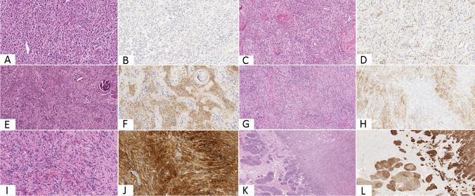
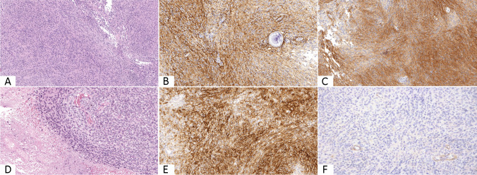
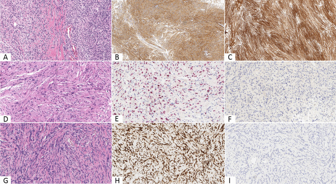
Meningiomas are common tumors of the central nervous system. Although their histological diagnosis is usually straightforward, their differential diagnosis versus other tumors may be challenging at times. The objective of this study is to assess the diagnostic value of CD13 immunoexpression in the differential diagnosis between meningiomas and their morphological mimics. Immunohistochemical analysis for CD13, epithelial membrane antigen, SOX10, and STAT6 was carried out in a large cohort of primary meningeal tumors comprising 225 meningiomas, 15 schwannomas, and 20 solitary fibrous tumor/hemangiopericytomas. Within the meningioma group, the expression of CD13 and epithelial membrane antigen was distinguished in three categories using a semiquantitative score. Most of meningiomas expressed CD13 (94%) and epithelial membrane antigen (96%) while none of the schwannomas nor of the solitary fibrous tumor/hemangiopericytomas was positive for either the two markers. Diffuse positivity for CD13 and epithelial membrane antigen was more common in low-grade meningiomas than in anaplastic ones, which were also more often negative for such markers, especially for CD13 (32%). CD13 is a helpful immunohistochemical marker for the differential diagnosis of meningiomas and their mimics, achieving in combination with epithelial membrane antigen maximal sensitivity (100%) and showing statistically relevant difference of expression in comparison with both schwannomas (p < 0.0001) and solitary fibrous tumor/hemangiopericytomas (p < 0.0001). Furthermore, loss of CD13 expression could be related to outcome as it is associated with worrisome histological findings, mainly in the setting of anaplastic meningiomas.
Keywords: CD13; Differential diagnosis; Immunohistochemistry; Meningeal tumors; Meningiomas.
© 2022. The Author(s).
Conflict of interest statement
The authors declare no competing interests.
Figures
References
-
- Perry A, Louis DN, Budka H et al (2016) Meningioma. In: Louis DN, Ohgaki H, Wiestler OD, Cavenee WK (eds) WHO classification of tumours of the central nervous system, 4th edn. IARC press, Lyon, France, pp 232–245
MeSH terms
Substances
LinkOut - more resources
Full Text Sources
Research Materials
Miscellaneous
