Review
. 2022 Apr;30(2):397-434.
doi: 10.1007/s10787-022-00931-1. Epub 2022 Feb 25.

Natural compounds as safe therapeutic options for ulcerative colitis

Affiliations
Review

Natural compounds as safe therapeutic options for ulcerative colitis

Mukta Gupta et al. Inflammopharmacology. 2022 Apr.

Abstract

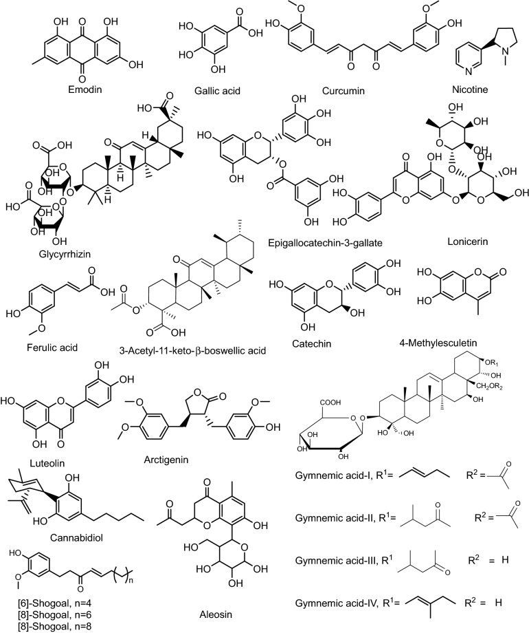
Ulcerative colitis (UC) is a chronic inflammatory bowel disease of unknown etiology. Several conventional treatments for UC such as corticosteroids, immunosuppressive agents, tumor necrosis factor antagonist, integrin blockers, and interleukin antagonist, and salicylates are available but are associated with the various limitations and side-effects. None of the above treatments helps to achieve the ultimate goal of the therapy, i.e., maintenance of remission in the long-term. Natural remedies for the treatment of UC show comparatively less side effects as compared to conventional approaches, and affordable. The current review presents details on the role of herbal drugs in the treatment and cure of UC. Google, PubMed, Web of Science, and Scopus portals have been searched for potentially relevant literature to get the latest developments and updated information related to use of natural drugs in the treatment of UC. Natural products have been used over centuries to treat UC. Some of the essential herbal constituents exhibiting antiulcerogenic activity include gymnemic acid (Gymnema sylvestre), shagoal (Zingiber officinale), catechin (Camellia sinensis), curcumin (Curcuma longa), arctigenin (Arctium lappa), and boswellic acid (Boswellia serrata). Although many plant-derived products have been recommended for UC, further research to understand the exact molecular mechanism is still warranted to establish their usefulness clinically.

Keywords: Anti-ulcerogenic activity; Herbal constituents; Inflammatory bowel disease; Ulcerative colitis.


Conflict of interest statement

Authors confirm that there are no known conflicts of interest associated with this work.

Figures

Fig. 1
Fig. 1
Chemical structures of bioactive constituents of herbal products posessing potential against UC
Fig. 2
Fig. 2
Photomicrographs of the rat colon stained with hematoxylin and eosin stain (× 40). Photomicrographs of protective A AV 50 mg/kg, B AV 300 mg/kg, C C+, D C−, and treatment groups, E AV 50 mg/kg, F AV 300 mg/kg, G C+, H C− in colitis rats. AV, Aloe vera (Bahrami et al. 2020)
Fig. 3
Fig. 3
Representative of the microscopic slide sections of the colon samples in rats: A Normal colon sample magnification (× 40), showed the normal structure of mucosa with an intact epithelial surface, submucosa, and muscular layer. B Normal colon sample, magnification (× 100), showed the normal structure of layers. C Acetic acid-induced colitis with no treatment, magnification (× 40), showed an inflammatory reaction, presence of ulcer, inflammation, edema, and diffuse infiltration of leukocytes in the submucosal layer. D Acetic acid-induced colitis with no treatment, magnification (× 200), showed inflammation in the submucosal layer with ulceration and edema. E Acetic acid-induced colitis + oral administration of sulfasalazine, magnification (× 40), revealed inflammatory reaction in the serosa layer. F Acetic acid-induced colitis + oral administration of sulfasalazine sample, magnification (× 100) revealed inflammatory reaction with the dominance of lymphocyte and fewer neutrophils with tissue necrosis. G, H Acetic acid induced colitis + oral administration of aloe vera gel, magnification (× 40), and (× 100), revealed the improvement of inflammation and the normal tissue (Hassanshahi et al. 2020)
Fig. 4
Fig. 4
Histological analysis of mice. A Colon section from control (water treated) animals showing normal colon tissue architecture (HE, × 400); B Colon section from Arctium lappa L. (AL)-treated animals showing normal histological architecture with no inflammatory cell infiltration, edema or crypt abscesses (HE, × 400); C, D Colon section from dextran sulfate sodium (DSS)-treated animals showing severe submucosal erosion with edema, ulceration, inflammatory cell infiltration (indicated with arrows) and crypt abscesses as well as epithelioglandular hyperplasia (C HE, × 200, D HE, × 400); E Colon section from animals challenged with DSS after prior treatment with AL showing normal histological architecture with slight inflammatory cell infiltration and no submucosal edema or abnormality of crypt cells (HE, × 400). All the results are representative of three independent experiments (Huang et al. 2010)
Fig. 5
Fig. 5
Effects of the ethanol extract of ALF on DSS-induced colitis in mice. Mice were treated with 3.5% DSS in drinking water for 7 days followed by drinking water for 3 days. ALE (25, 50, 100 mg/kg) were administrated orally once a day for 10 consecutive days. Mice were sacrificed at day 10. A DAI as the average score of body weight loss, stool consistency and rectal bleeding was scored from 0 to 4 (n = 7–8). B The colon length of each group at day 10 (n = 7–8). C The levels of MPO in colon (n = 7–8). D Histological changes of colon, characterized by distinct infiltration of inflammatory cells (arrow) and crypt destruction (solid circle) (magnification × 200). E Histological scores of colon from each group (n = 6). All data are presented as mean ± SEM. ##P < 0.01 vs. normal group, *P < 0.05, **P < 0.01 vs. DSS group. ALE ethanol extract of ALF, MES mesalazine (Wu et al. 2014)
Fig. 6
Fig. 6
a Hematoxylin and eosin stain (H&E), b Masson’s trichrome (MT), and c Periodic acid-Schiff (PA) staining sections of control group colon with normal intact cross section structure. Panel d shows the AKBA treated tissue sections of colon H&E stained. Panel e colon section with MT stained. Panel f shows colon section PA stained with maintained histological structure. In Panel g the yellow arrow shows the infiltration of mixed leucocytes in cross section and H&E stained DSS treated colon. h DSS treated MT stained colon section with yellow arrow showing the infiltration of inflammatory cells. Panel i shows the DSS treated colon, PA stained specifying the mixed inflammatory cells infiltration sites in the tissue section. Panels jl show the DSS + AKBA colon sections stained H&E, MT and PA. Yellow arrows represent the reduced infiltration of mixed inflammatory cells and well maintained tissue architecture (Roy et al. 2020)
Fig. 7
Fig. 7
ETO-curcumin exerts superior anti-inflammatory effects compared to standard curcumin on DSS-induced inflammation at 50 mg/kg body weight. A Graphical representation of curcumin treatment strategy. B Changes in individual categories of disease activity index (DAI) (top), body weight changes, stool consistency and stool bleeding (left–right). Changes in DAI (bottom). C Representative image of colons (left) and average colon length (right). D Spleen weight and E histology score on day 14. F Representative hematoxylin and eosin (H&E) staining of large intestine on day 14. × 100 magnification (left) and × 400 magnification (right). *P < 0.05, **P < 0.01, ***P < 0.001(Toden et al. 2017)
Fig. 8
Fig. 8
Treatment with Lico A relieved colonic microscopic damages in mice with DSS-induced UC. A Histological alteration of colonic mucosal. Histopathological sections were stained by H&E. Representative results are shown. Normal control (a); UC control (b) or Lico A, 20 mg/kg (c), 40 mg/kg (d) and 80 mg/kg (e). Original magnification × 200. B Histopathological scores of colons. Five specimens were selected randomly for histopathological study. Data are expressed as the means and individual histopathological scores, **P < 0.01 (Liu et al. 2018)
Fig. 9
Fig. 9
Histopathological sections of colons from rats stained with H&E (× 400). Colonic microscopic image of A normal rat colon from Cont group with intact mucosal layer and epithelial; B acetic acid (AA) treated rat colon with diffused active colitis, extensive damage including edema in submucosa and chronic inflammatory cells infiltrate with widely ulcerating mucosa, and hemorrhages; CE dose dependent reparative epithelial changes and ulcer healing with lymphoid follicle in colon of GS treated rats (50, 100 and 200 mg/kg, respectively); F attenuated cell damage with complete ulcer healing in MES treated group (Aleisa et al. 2014)
Fig. 10
Fig. 10
Microscopic study (original magnification × 50) of colons of mice with DSS-induced colitis treated with BuOH extracts of L. japonica. Treatment dose was 1 mg/kg, 10 mg/kg and 100 mg/kg, respectively, and 5-ASA was 100 mg/kg (Lee et al. 2011)
Fig. 11
Fig. 11
Effects of the water extract of Lonicera japonica (LJE) on histological findings and score in dextran sulphate sodium (DSS)-induced colitis. The images ae are the representative histological findings of all groups: a normal, b DSS, c DSS þ LJE 20 mg/kg, d DSS þ LJE 100 mg/kg and e DSS þ LJE 500 mg/kg groups, cryptal grand, surface of the epithelium;, neutrophils. The graph shows the histological scores. The score of the normal group is zero. Values are means (n = 8 for each group), with their standard errors represented by vertical bars. Mean values were significantly different compared with the DSS group: *P < 0·05, **P < 0·01 (Park et al. 2013)
Fig. 12
Fig. 12
Microscopic view and the total histological index score of the proximal colon of mice in all groups of the current study. a Group 1 (negative control): intact epithelium with normal epithelial cells infiltration (Sum score 0); b Group 2 (control + ve DSS exposure without treatment): focal epithelial ulceration (black arrows) with transmural infiltration of inflammatory cells (Sum score 5); c Group 3 (vehicle control group): intact epithelial surface with transmural infiltration of inflammatory cells (Sum score 4); d Group 4 (DSS exposure and 6-TG treatment): intact epithelial surface with moderate infiltration of inflammatory cells in mucosa and submucosa (Sum score 2); e Group 5 (DSS exposure and 20 mg/kg BW Shogaol treatment): intact epithelium with mild infiltration of inflammatory cells in mucosa only (Sum score 1); f Group 6 (DSS exposure and 40 mg/kg BW Shogaol treatment): intact epithelium with no inflammatory cells infiltration (Sum score 0). H&E stain; Black dash line indicated the extent of inflammatory cells infiltration; scale bar 100 μm. DSS dextran sodium sulfate, BW body weight, 6-TG 6-thioguanine (Hassan and Hassan 2018)
Fig. 13
Fig. 13
Microscopic view and the total histological index score of the distal colon of mice in all groups of the current study. a Group 1 (negative control): intact epithelium with normal epithelial cells infiltration (Sum score 0); b Group 2 (control + ve DSS exposure without treatment): extensive epithelial ulceration (black arrows) with transmural infiltration of inflammatory cells (Sum score 6); c Group 3 (vehicle control group): focal epithelial erosion (black arrow) with transmural infiltration of inflammatory cells (Sum score 4); d Group 4 (DSS exposure and 6-TG treatment): intact epithelial surface with moderate infiltration of inflammatory cells in mucosa and submucosa (Sum score 2); e Group 5 (DSS exposure and 20 mg/kg BW Shogaol treatment): intact epithelium with moderate infiltration of inflammatory cells in mucosa and submucosa (Sum score 2); f Group 6 (DSS exposure and 40 mg/kg BW Shogaol treatment): intact epithelium with mild infiltration of inflammatory cells in the mucosa (Sum score 1). H&E stain; Black dash line indicated the extent of inflammatory cells infiltration; scale bar 100 μm. DSS dextran sodium sulfate, BW body weight, 6-TG 6-thioguanine (Hassan and Hassan 2018)

References

    1. Aggarwal BB, Harikumar KB. Potential therapeutic effects of curcumin, the anti-inflammatory agent, against neurodegenerative, cardiovascular, pulmonary, metabolic, autoimmune and neoplastic diseases. Int J Biochem Cell Biol. 2009;41:40–59. doi: 10.1016/j.biocel.2008.06.010. - DOI - PMC - PubMed
    1. Akkol EK, Acıkara ÖB, Süntar I, Ergene B, Çitoğlu GS. Ethnopharmacological evaluation of some Scorzonera species: in vivo anti-inflammatory and antinociceptive effects. J Ethnopharmacol. 2012;140:261–270. doi: 10.1016/j.jep.2012.01.015. - DOI - PubMed
    1. Aleisa AM, Al Rejaie SS, Abuohashish HM, Ola MS, Parmar MY, Ahmed MM. Pretreatment of Gymnemasylvestre revealed the protection against acetic acid-induced ulcerative colitis in rats. BMC Complement Altern Med. 2014;14:49. doi: 10.1186/1472-6882-14-49. - DOI - PMC - PubMed
    1. Algieri F, Rodriguez-Nogales A, Rodriguez-Cabezas ME, Risco S, Ocete M, Galvez J. Botanical drugs as an emerging strategy in inflammatory bowel disease: a review. Mediat Inflamm. 2015 doi: 10.1155/2015/179616. - DOI - PMC - PubMed
    1. Al-Snafi A. The pharmacological importance and chemical constituents of Arctiumlappa. A review. Int J Pharm Res Scholar. 2014;3:663–670.

MeSH terms

LinkOut - more resources