. 2022 Jun;19(3):603-616.
doi: 10.1007/s13770-022-00435-7. Epub 2022 Feb 25.

Gymnemic Acid Ameliorates Pancreatic β-Cell Dysfunction by Modulating Pdx1 Expression: A Possible Strategy for β-Cell Regeneration

Affiliations

Gymnemic Acid Ameliorates Pancreatic β-Cell Dysfunction by Modulating Pdx1 Expression: A Possible Strategy for β-Cell Regeneration

Pugazhendhi Kannan et al. Tissue Eng Regen Med. 2022 Jun.

Abstract

Background: Endogenous pancreatic β-cell regeneration is a promising therapeutic approach for enhancing β-cell function and neogenesis in diabetes. Various findings have reported that regeneration might occur via stimulating β-cell proliferation, neogenesis, or conversion from other pancreatic cells to β-like cells. Although the current scenario illustrates numerous therapeutic strategies and approaches that concern endogenous β-cell regeneration, all of them have not been successful to a greater extent because of cost effectiveness, availability of suitable donors and rejection in case of transplantation, or lack of scientific evidence for many phytochemicals derived from plants that have been employed in traditional medicine. Therefore, the present study aims to investigate the effect of gymnemic acid (GA) on β-cell regeneration in streptozotocin-induced type 1 diabetic rats and high glucose exposed RIN5-F cells.

Methods: The study involves histopathological and immunohistochemical analysis to examine the islet's architecture. Quantitative polymerase chain reaction (qPCR) and/or immunoblot were employed to quantify the β-cell regeneration markers and cell cycle proliferative markers.

Results: The immunoexpression of E-cadherin, β-catenin, and phosphoinositide 3-kinases/protein kinase B were significantly increased in GA-treated diabetic rats. On the other hand, treatment with GA upregulated the pancreatic regenerative transcription factor viz. pancreatic duodenal homeobox 1, Neurogenin 3, MafA, NeuroD1, and β-cells proliferative markers such as CDK4, and Cyclin D1, with a simultaneous downregulation of the forkhead box O, glycogen synthase kinase-3, and p21cip1 in diabetic treated rats. Adding to this, we noticed increased nuclear localization of Pdx1 in GA treated high glucose exposed RIN5-F cells.

Conclusion: Our results suggested that GA acts as a potential therapeutic candidate for endogenous β-cell regeneration in treating type 1 diabetes.

Keywords: Diabetes; Gymnemic acid; Islet architecture; β-Cell proliferation; β-Cell regeneration.


Conflict of interest statement

The authors declare that they have no conflict of interest.

Figures
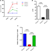
Fig. 1
Fig. 1
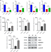
A Assessment of fasting blood glucose, B Plasma insulin levels and C Insulin mRNA expression levels in control and experimental groups. Data are shown as mean ± SEM, n = 6 for each group. Group 2 compared with Group 1; Group 3 compared with Group 2. Statistical significance represented with asterisks (*represents p < 0.05, **represents p < 0.01, ***represents p < 0.001, ns no significant)
Fig. 2
Fig. 2
A Pancreatic tissues histology of diabetic rats and treatment with GA (H & E × 40). Group 1 (G1)—Tissue section of control rats showed normal cellular architectures with the islet (dotted line) of Langerhans and healthy blood vessels. Group 2 (G2)—STZ induced diabetic rats showed severe vacuolation, degranulation, and massive destruction of the islets were observed (arrowheads). Group 3 (G3)—Diabetic rats supplemented with gymnemic acid showed significant improvement in cellular architecture with reduced degeneration of pancreatic islets. B Immunohistochemistry evaluation of insulin in pancreatic tissues of experimental rats. G1—Control rats showed the typical cellular architecture and positive insulin islets (dotted line). G2—Diabetic rats show a drastic decline of insulin content (arrowheads). G3—Diabetic rats treated with gymnemic shows increase of insulin content (arrowheads) and the moderate number of positive islets
Fig. 3
Fig. 3
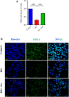
Pancreatic mRNA and protein expression levels of regeneration markers viz. Pdx1, Neurognin 3, NeuroD1, and MafA in GA treated diabetic rats. A Relative mRNA expression levels and B protein expression levels. Data are shown as mean ± SEM of three independent observations in each group. Group 2 compared with Group 1; Group 3 compared with Group 2. Statistical significance represented with asterisks (*represents p < 0.05, **represents p < 0.01, ***represents p < 0.001, ns no significant)
Fig. 4
Fig. 4
GA mediated activation of PI3K/AKT and cell cycle proliferative markers in various experimental groups. A Immunoblot analysis of E-cadherin, β-catenin, PI3K, AKT, pAKT and B FOXO1, GSK3β. C Cell cycle markers Cyclin D1, CDK4, and p21cip1. Data are shown as mean ± SEM of three independent observations in each group. Group 2 compared with Group 1; Group 3 compared with Group 2. Statistical significance represented with asterisks (*represents p < 0.05, **represents p < 0.01, ***represents p < 0.001, ns no significant)
Fig. 5
Fig. 5
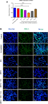
Effect of GA on insulin and Pdx1 in RIN-5F cells. A Insulin mRNA expression and B Cellular localization Pdx1 in RIN-5F cells treated with/without high glucose and GA treatment (Magnification 40X)
Fig. 6
Fig. 6
A The siRNA transfection efficiency analysis by qPCR. Cells were transfected with scrambled (Scr) siRNA and siRNA targeting Pdx1 for various experimental time (6 h, 12 h, 24 h, 48 h and 72 h). Statistical significance represented with asterisks (*represents p < 0.05, **represents p < 0.01, ***represents p < 0.001, ns no significant). B Influence of GA on RNAi mediated gene silencing of Pdx1 in RIN-5F cells. Cells were transfected with scrambled (Scr) siRNA and siRNA targeting Pdx1 for 24 h in the concentration of 30 pmol. Subcellular localization of Pdx1 in RNAi mediated transient knockdown in RIN-5F cells along with treated with/without high glucose and GA treatment (Magnification: 40×)

Similar articles

Cited by

References

    1. Zhu Y, Liu Q, Zhou Z, Ikeda Y. PDX1, Neurogenin-3, and MAFA: critical transcription regulators for beta cell development and regeneration. Stem Cell Res Ther. 2017;8:240. doi: 10.1186/s13287-017-0694-z. - DOI - PMC - PubMed
    1. Jiang WJ, Peng YC, Yang KM. Cellular signaling pathways regulating β-cell proliferation as a promising therapeutic target in the treatment of diabetes. Exp Ther Med. 2018;16:3275–3285. - PMC - PubMed
    1. Richardson T, Kerr D. Skin-related complications of insulin therapy: epidemiology and emerging management strategies. Am J Clin Dermatol. 2003;4:661–7. - PubMed
    1. Farney AC, Sutherland DE, Opara EC. Evolution of islet transplantation for the last 30 years. Pancreas. 2016;45:8–20. doi: 10.1097/MPA.0000000000000391. - DOI - PubMed
    1. Zhong F, Jiang Y. Endogenous pancreatic β cell regeneration: a potential strategy for the recovery of β cell deficiency in diabetes. Front Endocrinol. 2019;20:101. doi: 10.3389/fendo.2019.00101. - DOI - PMC - PubMed

Publication types

LinkOut - more resources