. 2022 Feb 25;17(2):e0263839.
doi: 10.1371/journal.pone.0263839. eCollection 2022.

Proteasome inhibition-enhanced fracture repair is associated with increased mesenchymal progenitor cells in mice

Affiliations

Proteasome inhibition-enhanced fracture repair is associated with increased mesenchymal progenitor cells in mice

Hengwei Zhang et al. PLoS One. .

Abstract

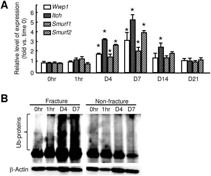
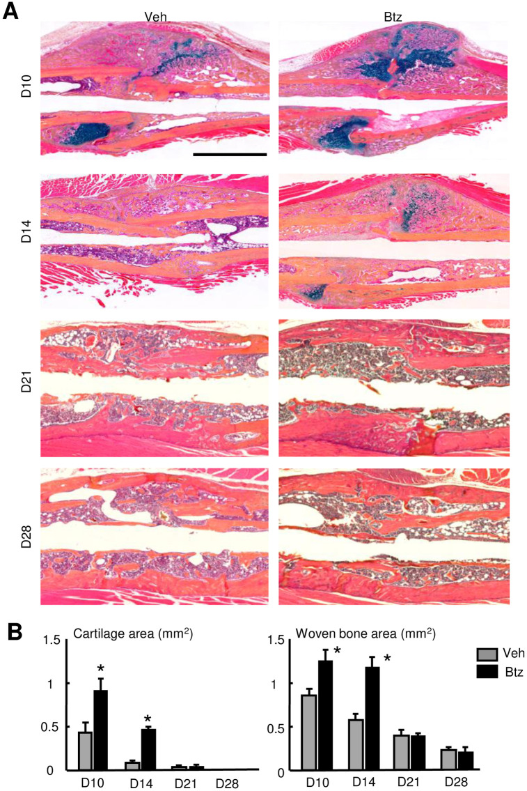
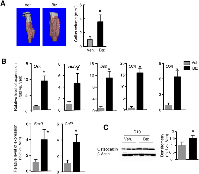
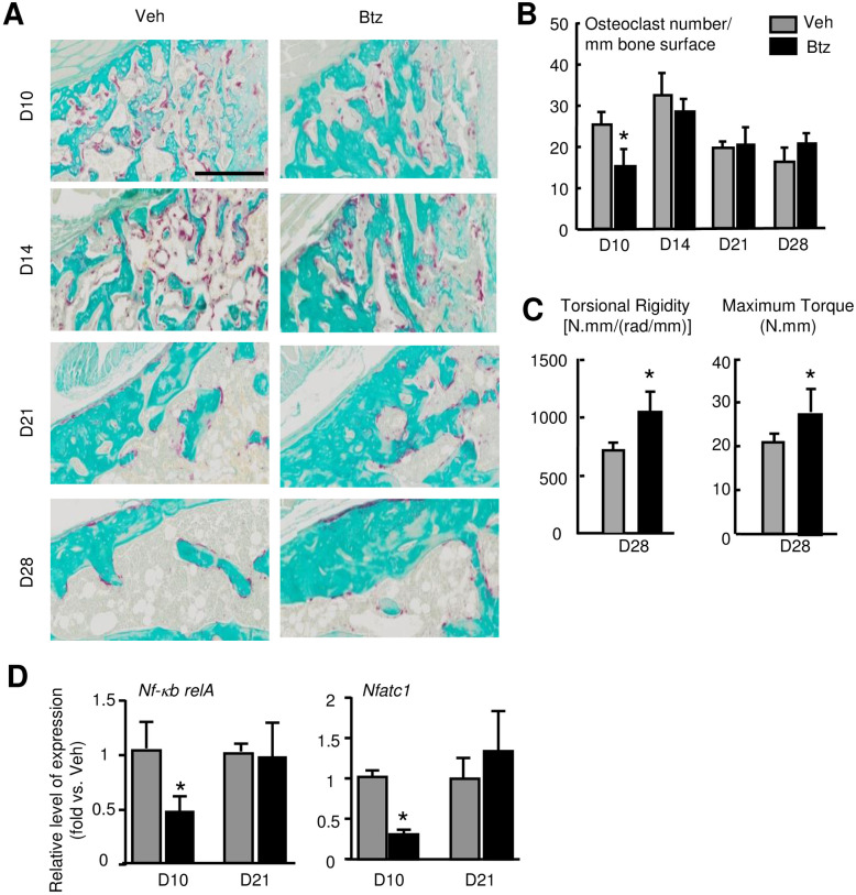
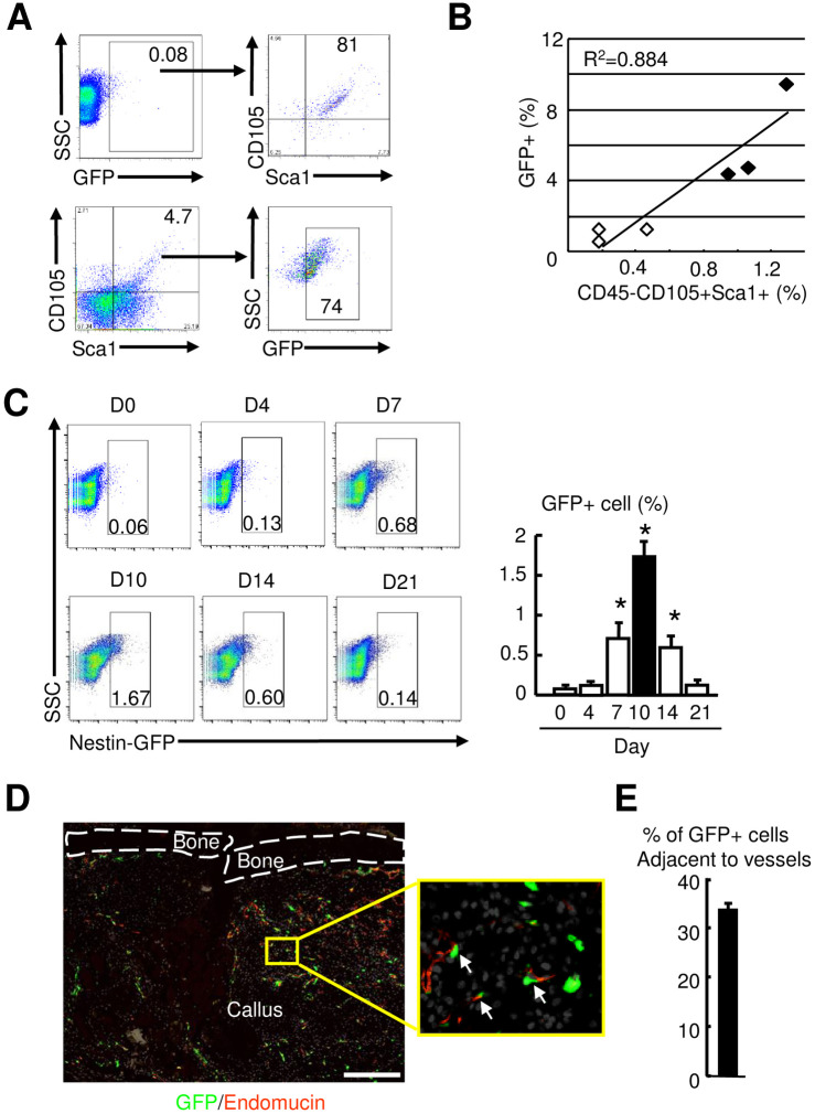
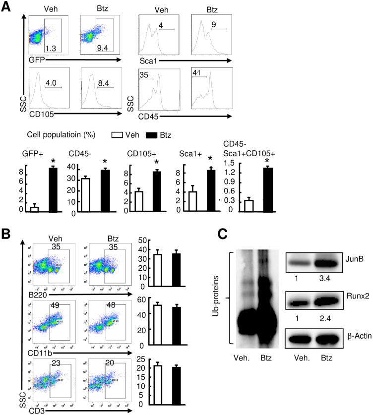
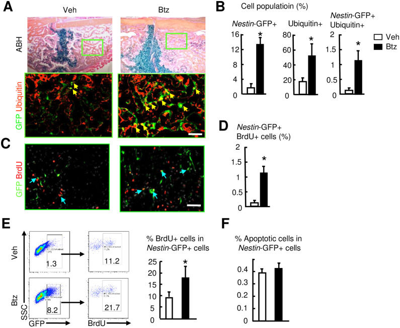
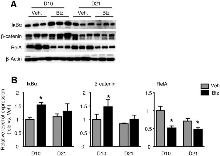
The ubiquitin/proteasome system controls the stability of Runx2 and JunB, proteins essential for differentiation of mesenchymal progenitor/stem cells (MPCs) to osteoblasts. Local administration of proteasome inhibitor enhances bone fracture healing by accelerating endochondral ossification. However, if a short-term administration of proteasome inhibitor enhances fracture repair and potential mechanisms involved have yet to be exploited. We hypothesize that injury activates the ubiquitin/proteasome system in callus, leading to elevated protein ubiquitination and degradation, decreased MPCs, and impaired fracture healing, which can be prevented by a short-term of proteasome inhibition. We used a tibial fracture model in Nestin-GFP reporter mice, in which a subgroup of MPCs are labeled by Nestin-GFP, to test our hypothesis. We found increased expression of ubiquitin E3 ligases and ubiquitinated proteins in callus tissues at the early phase of fracture repair. Proteasome inhibitor Bortezomib, given soon after fracture, enhanced fracture repair, which is accompanied by increased callus Nestin-GFP+ cells and their proliferation, and the expression of osteoblast-associated genes and Runx2 and JunB proteins. Thus, early treatment of fractures with Bortezomib could enhance the fracture repair by increasing the number and proliferation of MPCs.


Conflict of interest statement

NO authors have competing interests.

Figures

Fig 1
Fig 1. Increased expression of E3 ligases and ubiquitinated proteins in fracture callus.
3-month-old C57BL/6J male mice received an open tibial fracture surgery. (A) Expression levels of E3 ligases in callus tissues determined by qPCR at different time points. N = 3 mice. Values are means ± SD. Relative mRNA expression is the fold-change vs. the time 0 samples as 1. *p<0.05 vs. time 0 samples. (B) Total Ub-proteins in callus tissues determined by Western blot analysis using anti-Ubiquitin Ab at different time points. N = 3 mice. Experiments were repeated independently at least 2 times.
Fig 2
Fig 2. Bortezomib increases cartilage and woven bone areas in fracture callus.
3-month-old C57BL/6J male mice received fracture surgery as in Fig 1. Mice were treated with Btz (0.6 mg/kg body weight, i.p.) on 1-, 3- and 5-dpf, and were sacrificed at different times. (A) Representative ABH-stained sections showing higher woven bone and callus areas in Btz-treated mice. Scale bar = 1 mm. (B) Woven bone and cartilage areas analyzed by Image J software. N = 8 mice. Values are means ± SD. *p< 0.05 vs. vehicle-treated mice.
Fig 3
Fig 3. Bortezomib increases callus volume and the expression of genes related to osteoblast and chondrocyte differentiation in fracture callus.
3-month-old C57BL/6J male mice received fracture surgery and treatment as in Fig 2. (A) Callus volume measured at 10 dpf by micro-CT. N = 8 mice. Values are means ± SD. *p< 0.05 vs. vehicle-treated mice. (B) Expression levels of genes related to OB differentiation and maturation, and chondrocyte differentiation in callus tissues examined by qPCR at 10 dpf. N = 3 mice. Values are means ± SD. Relative mRNA expression is the fold-change vs. vehicle-treated mice as 1. *p< 0.05 vs. vehicle-treated mice. (C) Expression levels of Osteocalcin in callus tissues following Btz treatment at 10 dpf examined by Western blot analysis. Data is normalized to callus samples from vehicle-treated mice. N = 3 mice. *p< 0.05 vs. vehicle-treated mice.
Fig 4
Fig 4. Bortezomib decreases osteoclasts and improves fracture repair in mice.
3-month-old C57BL/6J male mice received fracture surgery were treated with Btz as in Fig 2. (A) Representative TRAP-stained sections showing fewer TRAP+ osteoclasts at 10 dpf in Btz-treated mice. Scale bar = 0.25 mm. (B) TRAP+ osteoclasts in callus analyzed by Image J software. N = 8 mice. Values are means ± SD. *p< 0.05 vs. vehicle-treated mice. (C) Bone stiffness and strength assessed by biomechanical testing at 28 dpf. N = 5 mice. Values are means ± SD. *p< 0.05 vs. vehicle-treated mice. (D) Expression of osteoclast transcription factors, nf-kb and nfatc1 examined by qPCR at 10 dpf. N = 3. Values are means ± SD. Relative mRNA expression is the fold-change vs. samples from the vehicle-treated mice as 1. Unpaired t-test is used to compare Veh and Btz treated day 10 and day 21 callus, respectively. *p< 0.05 vs. vehicle-treated mice.
Fig 5
Fig 5. Increased Nestin-GFP+ mesenchymal stem/progenitor cells in callus at the early phase of fracture repair.
3-month-old Nestin-GFP male mice were used. (A) cells were stained with anti-CD105 and Sca1 Abs and subjected to flow cytometry. GFP+ (upper) or CD105+Sca1+ cells were gated for analysis. N = 5 mice. SSC: side-scattered light. (B) Correlation of Nestin-GFP+ and CD45-CD105+Sca1+ cells. (C) The percentage of Nestin-GFP+ cells in callus tissues analyzed by flow cytometry. N = 3. Values are means ± SD. *p< 0.05 vs. time 0 samples. (D) Frozen sections subjected to immunofluorescence stained with anti-Endomucin Ab for blood vessels. The insert indicates enlarged immunofluorescence stained image showing Nestin-GFP+ cells adjacent to Endomucin+ blood vessels (arrows). Scale bar = 1 mm. (E) The Nestin-GFP+ cells adjacent to Endomucin+ blood vessels (distance < 2 cell diameter to blood vessel) counted. The percentage of Nestin-GFP+ cells adjacent to blood vessels over the total number Nestin-GFP+ cells calculated. N = 3 mice. Values are means ± SD.
Fig 6
Fig 6. Bortezomib increases the number of Nestin-GFP+ cells and expression of osteogenic proteins in fracture callus.
3-month-old Nestin-GFP male mice received fracture surgery were treated with Btz as in Fig 2. Callus tissues were harvested at 10 dpf. The percentage of mesenchymal (A) and hematopoietic (B) lineage cell populations analyzed by flow cytometry. N = 4 mice. Values are means ± SD. *p< 0.05 vs. vehicle-treated mice. (C) Expression levels of total Ub-proteins, Runx2, and JunB examined by Western blot analysis. Numbers are the fold-change by densitometry analysis of Western blot images using callus samples harvested at 10 dpf from vehicle-treated mice as 1. N = 3 mice.
Fig 7
Fig 7. Bortezomib increases the proliferation of Nestin-GFP+ cells in fracture callus.
3-month-old Nestin-GFP male mice received fracture surgery were treated with Btz as in Fig 2. Callus tissues were harvested at 10 dpf. (A) Frozen sections subjected to immunofluorescence staining with anti-Ubiquitin Ab and examined under a fluorescent microscopy. Nestin-GFP+ cells are green and Ubiquitin+ cells are stained for red. Nestin-GFP+Ubiquitin+ cells are stained for yellow (indicated by yellow arrows). Scale bar = 1 mm. The adjacent sections stained for ABH. Inserts indicate enlarged immunofluorescence stained images. (B) The percentage of Nestin-GFP+, Ubiquitin+, or Nestin-GFP+Ubiquitin+ cells were counted. N = 3 mice. Values are means ± SD. *p< 0.05 vs. vehicle-treated mice. (C) Mice were given two injections of BrdU (1 mg/i.p. injection) spaced 16 hours apart, starting at 9 dpf. Frozen sections subjected to immunofluorescence staining with anti-BrdU Ab and analyzed under a fluorescent microscopy. Nestin-GFP+ cells are green and BrdU+ cells are stained for red. Nestin-GFP+BrdU+ cells are stained for yellow (indicated yellow arrows). Scale bar = 1 mm. (F) The percentage of Nestin-GFP+BrdU+ cells was counted. N = 3 mice. Values are means ± SD. *p< 0.05 vs. vehicle-treated mice. (D) The percentage of BrdU+ cells in callus Nestin-GFP+ cells analyzed by flow cytometry. N = 3 mice. Values are means ± SD. *p< 0.05 vs. vehicle-treated mice. (E) Cell apoptosis assessed by flow cytometry as the percentage of Annexin V+ cells in Nestin-GFP+ cells. N = 3 mice. Values are means ± SD.
Fig 8
Fig 8. Bortezomib increases IκBα, β-catenin and decreases RelA protein levels in fracture callus at 10-day post-fracture.
3-month-old C57BL/6J mice received fracture surgery were treated with Btz as in Fig 2. Callus tissues were harvested at 10 and 21 dpf. (A) Expression levels of IκBα, β-catenin and RelA examined by Western blot analysis. (B) Densitometry analysis of Western blot images in A. Data is normalized to 10 dpf callus samples from vehicle-treated mice. N = 3 mice. Unpaired t-test is used to compare vehicle- and Btz-treated 10 dpf and 21 dpf callus, respectively. *p< 0.05 vs. vehicle-treated mice.

Similar articles

Cited by

References

    1. Clark D, Nakamura M, Miclau T, Marcucio R. Effects of Aging on Fracture Healing. Curr Osteoporos Rep. 2017;15(6):601–8. Epub 2017/11/17. doi: 10.1007/s11914-017-0413-9 . - DOI - PMC - PubMed
    1. Curtis EM, van der Velde R, Moon RJ, van den Bergh JP, Geusens P, de Vries F, et al.. Epidemiology of fractures in the United Kingdom 1988–2012: Variation with age, sex, geography, ethnicity and socioeconomic status. Bone. 2016;87:19–26. Epub 2016/03/13. doi: 10.1016/j.bone.2016.03.006 . - DOI - PMC - PubMed
    1. Pountos I, Georgouli T, Henshaw K, Bird H, Jones E, Giannoudis PV. The effect of bone morphogenetic protein-2, bone morphogenetic protein-7, parathyroid hormone, and platelet-derived growth factor on the proliferation and osteogenic differentiation of mesenchymal stem cells derived from osteoporotic bone. J Orthop Trauma. 2010;24(9):552–6. Epub 2010/08/26. doi: 10.1097/BOT.0b013e3181efa8fe . - DOI - PubMed
    1. Morgan EF, Mason ZD, Bishop G, Davis AD, Wigner NA, Gerstenfeld LC, et al.. Combined effects of recombinant human BMP-7 (rhBMP-7) and parathyroid hormone (1–34) in metaphyseal bone healing. Bone. 2008;43(6):1031–8. Epub 2008/09/02. doi: 10.1016/j.bone.2008.07.251 . - DOI - PubMed
    1. Liu YC. Ubiquitin ligases and the immune response. Annual review of immunology. 2004;22:81–127. Epub 2004/03/23. doi: 10.1146/annurev.immunol.22.012703.104813 . - DOI - PubMed

Publication types

MeSH terms