. 2022 Aug 11;140(6):571-593.
doi: 10.1182/blood.2020006479.

Anemia in the pediatric patient

Affiliations

Anemia in the pediatric patient

Patrick G Gallagher. Blood. .

Abstract

The World Health Organization estimates that approximately a quarter of the world's population suffers from anemia, including almost half of preschool-age children. Globally, iron deficiency anemia is the most common cause of anemia. Other important causes of anemia in children are hemoglobinopathies, infection, and other chronic diseases. Anemia is associated with increased morbidity, including neurologic complications, increased risk of low birth weight, infection, and heart failure, as well as increased mortality. When approaching a child with anemia, detailed historical information, particularly diet, environmental exposures, and family history, often yield important clues to the diagnosis. Dysmorphic features on physical examination may indicate syndromic causes of anemia. Diagnostic testing involves a stepwise approach utilizing various laboratory techniques. The increasing availability of genetic testing is providing new mechanistic insights into inherited anemias and allowing diagnosis in many previously undiagnosed cases. Population-based approaches are being taken to address nutritional anemias. Novel pharmacologic agents and advances in gene therapy-based therapeutics have the potential to ameliorate anemia-associated disease and provide treatment strategies even in the most difficult and complex cases.


Figures

None
Graphical abstract
Figure 1.
Figure 1.
Mean Hb concentrations by age and sex (−2 SD values). Compiled from data from the United States, Europe, and White populations. Used with permission from Chaparro and Suchdev.
Figure 2.
Figure 2.
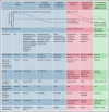
An approach to anemia based on mean corpuscular volume and reticulocyte count.
Figure 3.
Figure 3.
Anemia by age.
Figure 4.
Figure 4.
PB smears. PB smears from (A) hereditary spherocytosis. Dense, spherical-shaped erythrocytes are seen. (B) β-Thalassemia major. Hypochromic, microcytic erythrocytes, anisocytosis, and a nucleated red blood cell are seen. (C) Sideroblastic anemia. Polychromasia, anisopoikilocytosis, and basophilic stippling are seen in a case of X-linked congenital sideroblastic anemia. (D) Thrombotic thrombocytopenic purpura. Anisopoikilocytosis and marked schistocytosis are seen on the smear of an infant with Upshaw-Schulman syndrome. (E) Iron deficiency. Significant anisocytosis, hypochromia, and microcytosis are seen. (F) Vitamin B12 deficiency. Macro-ovalocytes, microcytes, and hypersegmented neutrophils are seen. Erythrocyte basophilic stippling is shown in inset. These images were originally published in the ASH Image Bank. (A) Teresa Scordino, Hereditary spherocytosis, 2016, #00060308. (B) Girish Venkataraman, β-thalassemia major, 2018, #00062081; (C) Katherine Calvo, Congenital sideroblastic anemia peripheral blood, 2015; #00060064; (D) Helle Borgstrøm Hager and Mari Tjernsmo Andersen, Thrombotic thrombocytopenic purpura, #00061402; (E) Iron deficiency anemia moderate, 2015, #00060219. (F) Volodymyr Shponka and Maria Proytcheva, Megaloblastic anemia caused by severe B12 deficiency in a breastfed infant. 2017, #00061082. © The American Society of Hematology.
Figure 5.
Figure 5.
Markers of iron deficiency. *Diagnostic thresholds for reticulocyte Hb content vary between the type of blood cell analyzer as well as for hepcidin and soluble transferrin receptor assays. Reprinted with permission from Pasricha et al.
Figure 6.
Figure 6.
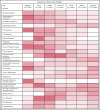
Features of hypochromic anemias.
Figure 7.
Figure 7.
BM examination. (A) Diamond-Blackfan anemia. BM aspirate shows a marked myeloid predominance. (B) Acquired aplastic anemia. High-power image of a hematoxylin and eosin-stained BM biopsy section from a teenage girl showing profound hypocellularity. The few remaining hematopoietic cells are lymphocytes, plasma cells, and macrophages with pigment in their cytoplasm. (C) Congenital dyserythropoietic anemia (CDA) type II. BM aspirate shows binucleated and multinucleated erythroid precursors. (D) Neuroblastoma. BM biopsy shows clumps of metastatic neuroblastoma cells. These images were originally published in the ASH Image Bank. (A) Amy Duffield, Diamond Blackfan Anemia Aspirate, 2015, #00060078. (B) Kristian T. Schafernak, Acquired Aplastic Anemia, 2016, #00060876. (C) Kristian T. Schafernak, Congenital Dyserythropoietic Anemia, Type II, 2016, #00060891. (D) Suzanne Vercauteren. Neuroblastoma Bone Marrow, 2015, #00060131. © The American Society of Hematology.

References

    1. Chaparro CM, Suchdev PS. Anemia epidemiology, pathophysiology, and etiology in low- and middle-income countries. Ann N Y Acad Sci. 2019;1450(1):15-31. - PMC - PubMed
    1. Zierk J, Hirschmann J, Toddenroth D, et al. Next-generation reference intervals for pediatric hematology. Clin Chem Lab Med. 2019;57(10):1595-1607. - PubMed
    1. Henry E, Christensen RD. Reference intervals in neonatal hematology. Clin Perinatol. 2015;42(3):483-497. - PubMed
    1. Ocas-Córdova S, Tapia V, Gonzales GF. Hemoglobin concentration in children at different altitudes in Peru: proposal for [Hb] correction for altitude to diagnose anemia and polycythemia. High Alt Med Biol. 2018;19(4):398-403. - PMC - PubMed
    1. Timmer T, Tanck MWT, Huis In ’t Veld EMJ, et al. Associations between single nucleotide polymorphisms and erythrocyte parameters in humans: a systematic literature review. Mutat Res Rev Mutat Res. 2019;779:58-67. - PubMed