. 2022 Oct 3;24(10):1673-1686.
doi: 10.1093/neuonc/noac054.

A preclinical model of patient-derived cerebrospinal fluid circulating tumor cells for experimental therapeutics in leptomeningeal disease from melanoma

Affiliations

A preclinical model of patient-derived cerebrospinal fluid circulating tumor cells for experimental therapeutics in leptomeningeal disease from melanoma

Vincent Law et al. Neuro Oncol. .

Erratum in

Abstract

Background: Leptomeningeal disease (LMD) occurs as a late complication of several human cancers and has no rationally designed treatment options. A major barrier to developing effective therapies for LMD is the lack of cell-based or preclinical models that recapitulate human disease. Here, we describe the development of in vitro and in vivo cultures of patient-derived cerebrospinal fluid circulating tumor cells (PD-CSF-CTCs) from patients with melanoma as a preclinical model to identify exploitable vulnerabilities in melanoma LMD.

Methods: CSF-CTCs were collected from melanoma patients with melanoma-derived LMD and cultured ex vivo using human meningeal cell-conditioned media. Using immunoassays and RNA-sequencing analyses of PD-CSF-CTCs, molecular signaling pathways were examined and new therapeutic targets were tested for efficacy in PD-CSF-CTCs preclinical models.

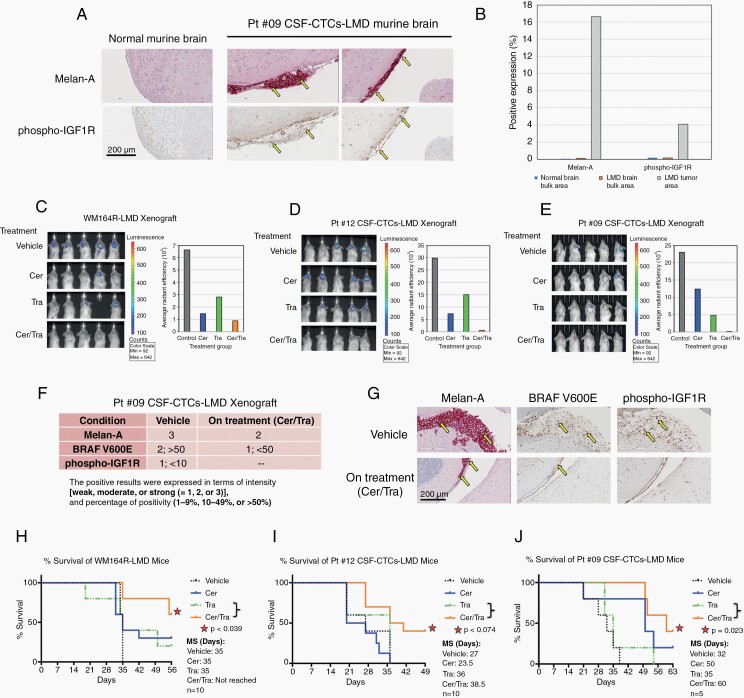
Results: PD-CSF-CTCs were successfully established both in vitro and in vivo. Global RNA analyses of PD-CSF-CTCs revealed several therapeutically tractable targets. These studies complimented our prior proteomic studies highlighting IGF1 signaling as a potential target in LMD. As a proof of concept, combining treatment of ceritinib and trametinib in vitro and in vivo demonstrated synergistic antitumor activity in PD-CSF-CTCs and BRAF inhibitor-resistant melanoma cells.

Conclusions: This study demonstrates that CSF-CTCs can be grown in vitro and in vivo from some melanoma patients with LMD and used as preclinical models. These models retained melanoma expression patterns and had signaling pathways that are therapeutically targetable. These novel models/reagents may be useful in developing rationally designed treatments for LMD.

Keywords: ceritinib; leptomeningeal disease (LMD); melanoma; patient-derived CSF-CTCs (PD-CSF-CTCs); single-cell RNA sequencing.


Figures

Figure 1.
Figure 1.
Ex vivo culture of melanoma PD-CSF-CTCs and examples of successful in vivo culture in LMD mouse model. (A) Immunoblot from a Human-Growth-Factor array comparing serum free medium, normal growth medium, HMC-conditioned medium, and HMC-conditioned medium that was cultured with CSF-CTCs. Arrows are pointing at duplicate blots representing each growth factor. (B) Representative bright field images of propagating ex vivo melanoma CSF-CTCs from four patients; three were BRAF V600E mutants (patients 9, 12, 16), and one was NRAS mutant (Pt #13). Each CSF-CTC culture displayed distinctive cell morphology. Bar = 100 μm. (C) Mutation status of growing PD-CSF-CTCs from patients 9 and 12 were evaluated by using single-nucleotide polymorphism genotyping. Both melanoma patients were BRAF V600E. HMC was used as control. (D) Representative immunofluorescence images showing that PD-CSF-CTCs are melanocytic in origin and expressed Melan-A, and α-SMA, whereas our negative controls (HMCs and human fibroblast cells FF2504) did not. Bar = 50 μm. (E) A schematic of how ex vivo CSF-CTCs were expanded. CTCs in CSF were expanded in vitro until sufficient cells were available to inoculate into a murine PDX and/or CDX models in vivo. CSF-CTCs were collected from LMD mice. (F) Brain MRIs of LMD mice from CDX model showed enlarged ventricles and hydrocephaly (arrows). G) H&E stained brain sections of patients 9 and 12 CDX LMD mice. Cancer cells metastasized in the meninges (arrows). Bar = 200 μm.
Figure 2.
Figure 2.
Transcriptome analysis of PD-CSF-CTCs shows adaptation to ex vivo culture and retention of cardinal melanoma genes including IGF1R, ErbB3, and Sox9. (A) t-SNE plot showing major cell types identified in patients 9’s and 12’s CSF-CTCs, and their respective in vitro and in vivo propagated PD-CSF-CTCs. (B) Venn diagrams showing the number of genes expressed that were unique to noncultured CSF-CTCs (Pt CSF) and those that were retained after in vitro (In vitro) and in vivo (In vivo) propagations. (C) Graph representing a list of 30 most enriched (logFC > 0.4 in gene expression; P < .05) melanoma-associated gene signatures that were retained after in vitro and in vivo propagations. (D) A list of 20 most enriched biological pathways in accordance to gene signatures in (C), identified by using the KEGG pathway database (E) Average log expression of melanoma-associated gene signatures of non-tumor cells (fibroblasts and immunocytes) in CSF and CSF-CTCs from LMD patients. Comparisons were made between patients 8, 9, 10, 11, and 12. (I) IHC scores of Melan-A, BRAF V600E, and phospho-IGF1R expression in brain and spinal cord tissues from patients 9 and 12, which were obtained from autopsies. (J) Representative autopsy slides of (I); Melan-A, BRAF V600E, and phospho-IGF1R staining by IHC. Bar = 200 μm.
Figure 3.
Figure 3.
IGF1R inhibition promotes cell growth inhibition. (A & B) CellTiter-Glo Assay was used to assess the effects of ceritinib and trametinib on growth inhibition at 72 hours in patients 9 and 12 PD-CSF-CTCs. Synergy between (Mean CI) ceritinib and trametinib was analyzed where values < 1 indicated synergism, and >1 indicated antagonism. (C) Immunoblot from a Proteome-Profiler-Human-Phospho-RTK array for patient 12 PD-CSF-CTCs after 24 hours ceritinib, trametinib, or both treatments. DMSO was used as control. Arrows are pointing at duplicate spots representing activities of phospho-IGF1R and -INSR. Average pixel intensity was quantified using ImageJ Software. (D) Schematic diagram of CRISPR/Cas9-mediated IGF1R deletion, and the incorporation of GFP for positive selection. (E) Representative 20x bright field and fluorescent images of patients 9 and 12 PD-CSF-CTCs, and HMC after 24 hours of CRSPR/Cas9-guided IGF1R deletion. GFP was used a positive marker. Bar = 400 μm. (F) Representative 20x bright field and fluorescent images showing transfection of GFP into PD-CSF-CTCs using lentiviral vector did not induce cell death after 48 hours. G) Average number of viable patients 9 and 12 PD-CSF-CTCs, and HMCs after 48 hours of IGF1R depletion.
Figure 4.
Figure 4.
Ceritinib + trametinib treatment prolonged survival of melanoma-associated LMD in vivo in murine xenograft model. (A) Representative images of Melan-A and phospho-IGF1R staining of mice normal and LMD brains by IHC. LMD mice were generated using PD-CSF-CTCs. Bar = 200 μm. (B) Quantitative analysis of Melan-A and phospho-IGF1R expressions in the brain sections of (A). LMD in mice were generated by inoculating (C) WM164R, (D) patient 12 PD-CSF-CTCs, and (E) patient 9 PD-CSF-CTCs in CSF. Disease progression in each cohort was assessed by BLI, after three weeks of ceritinib, trametinib, or both oral therapies. (F) IHC scores of Melan-A, BRAF V600E, and phospho-IGF1R expression in LMD xenograft brain tissues after three weeks of ceritinib and trametinib treatment. (G) Representative IHC sections of (F); Bar = 200 μm. Survival graphs of (H) WM164R-LMD, (I) patient 12 PD-CSF-CTCs-LMD, and (J) patient 9 PD-CSF-CTCs-LMD mice after receiving ceritinib, trametinib, or both treatments were analyzed. Control mice received vehicle solution. Median survival (MS) was assessed. Survival graphs were compared statistically using the Mantel-Cox test, performed by GraphPad Prism 6 Software.

Comment in

Similar articles

Cited by

References

    1. Glitza IC, Smalley KSM, Brastianos PK, et al. Leptomeningeal disease in melanoma patients: an update to treatment, challenges, and future directions. Pigment Cell Melanoma Res 2020;33(4):527–541. - PMC - PubMed
    1. Smalley I, Law V, Wyatt C, et al. Proteomic analysis of CSF from patients with leptomeningeal melanoma metastases identifies signatures associated with disease progression and therapeutic resistance. Clin Cancer Res. 2020;26(9):2163–2175. - PMC - PubMed
    1. Larkin J, Chiarion-Sileni V, Gonzalez R, et al. Five-year survival with combined nivolumab and ipilimumab in advanced melanoma. N Engl J Med. 2019;381(16):1535–1546. - PubMed
    1. Boire A, Zou Y, Shieh J, et al. Complement component 3 adapts the cerebrospinal fluid for leptomeningeal metastasis. Cell 2017;168(6):1101–1113.e13. - PMC - PubMed
    1. Chi Y, Remsik J, Kiseliovas V, et al. Cancer cells deploy lipocalin-2 to collect limiting iron in leptomeningeal metastasis. Science 2020;369(6501):276–282. - PMC - PubMed

Publication types