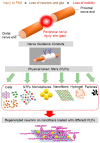
Advances in Electrospun Nerve Guidance Conduits for Engineering Neural Regeneration
- PMID: 35213952
- PMCID: PMC8876219
- DOI: 10.3390/pharmaceutics14020219
Advances in Electrospun Nerve Guidance Conduits for Engineering Neural Regeneration
Abstract
Injuries to the peripheral nervous system result in devastating consequences with loss of motor and sensory function and lifelong impairments. Current treatments have largely relied on surgical procedures, including nerve autografts to repair damaged nerves. Despite improvements to the surgical procedures over the years, the clinical success of nerve autografts is limited by fundamental issues, such as low functionality and mismatching between the damaged and donor nerves. While peripheral nerves can regenerate to some extent, the resultant outcomes are often disappointing, particularly for serious injuries, and the ongoing loss of function due to poor nerve regeneration is a serious public health problem worldwide. Thus, a successful therapeutic modality to bring functional recovery is urgently needed. With advances in three-dimensional cell culturing, nerve guidance conduits (NGCs) have emerged as a promising strategy for improving functional outcomes. Therefore, they offer a potential therapeutic alternative to nerve autografts. NGCs are tubular biostructures to bridge nerve injury sites via orienting axonal growth in an organized fashion as well as supplying a supportively appropriate microenvironment. Comprehensive NGC creation requires fundamental considerations of various aspects, including structure design, extracellular matrix components and cell composition. With these considerations, the production of an NGC that mimics the endogenous extracellular matrix structure can enhance neuron-NGC interactions and thereby promote regeneration and restoration of function in the target area. The use of electrospun fibrous substrates has a high potential to replicate the native extracellular matrix structure. With recent advances in electrospinning, it is now possible to generate numerous different biomimetic features within the NGCs. This review explores the use of electrospinning for the regeneration of the nervous system and discusses the main requirements, challenges and advances in developing and applying the electrospun NGC in the clinical practice of nerve injuries.
Keywords: extracellular matrix; fibrous scaffold; neural tissue engineering; peripheral nervous system; physical lumen filler; scaffold topography; structural support.
Conflict of interest statement
The authors confirm that there are no known conflicts of interest associated with this publication and there has been no significant financial support of this work that could have influenced its outcome.
Figures


References
-
- Zarrintaj P., Zangene E., Manouchehri S., Amirabad L.M., Baheiraei N., Hadjighasem M.R., Farokhi M., Ganjali M.R., Walker B.W., Saeb M.R., et al. Conductive biomaterials as nerve conduits: Recent advances and future challenges. Appl. Mater. Today. 2020;20:100784. doi: 10.1016/j.apmt.2020.100784. - DOI
Publication types
LinkOut - more resources
Full Text Sources

