Review
. 2022 Feb 4;14(2):359.
doi: 10.3390/pharmaceutics14020359.

Poly(Lactic Acid)-Based Microparticles for Drug Delivery Applications: An Overview of Recent Advances

Affiliations
Review

Poly(Lactic Acid)-Based Microparticles for Drug Delivery Applications: An Overview of Recent Advances

Antonios Vlachopoulos et al. Pharmaceutics. .

Abstract

The sustained release of pharmaceutical substances remains the most convenient way of drug delivery. Hence, a great variety of reports can be traced in the open literature associated with drug delivery systems (DDS). Specifically, the use of microparticle systems has received special attention during the past two decades. Polymeric microparticles (MPs) are acknowledged as very prevalent carriers toward an enhanced bio-distribution and bioavailability of both hydrophilic and lipophilic drug substances. Poly(lactic acid) (PLA), poly(lactic-co-glycolic acid) (PLGA), and their copolymers are among the most frequently used biodegradable polymers for encapsulated drugs. This review describes the current state-of-the-art research in the study of poly(lactic acid)/poly(lactic-co-glycolic acid) microparticles and PLA-copolymers with other aliphatic acids as drug delivery devices for increasing the efficiency of drug delivery, enhancing the release profile, and drug targeting of active pharmaceutical ingredients (API). Potential advances in generics and the constant discovery of therapeutic peptides will hopefully promote the success of microsphere technology.

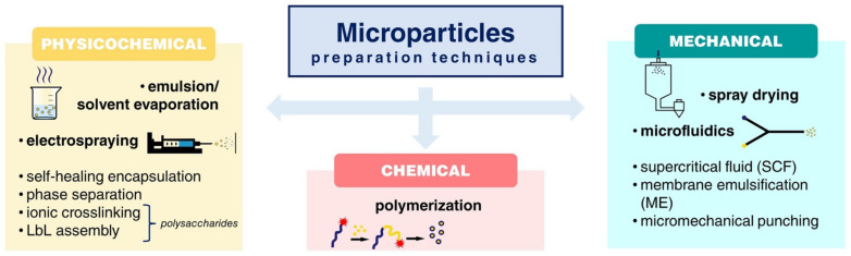
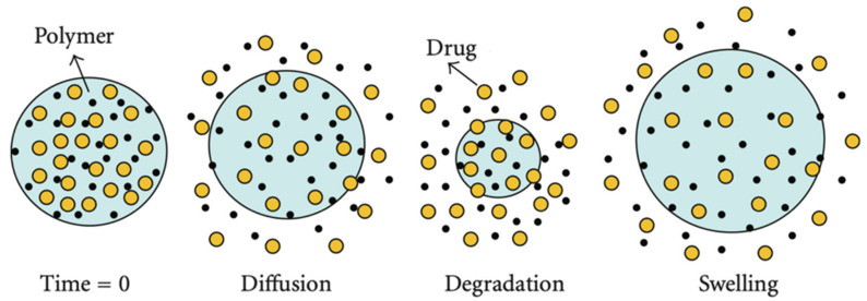
Keywords: copolymers; drug delivery; drug release mechanisms; microparticles; poly(lactic acid); preparation techniques.


Conflict of interest statement

The authors declare no conflict of interest. The company had no role in the design of the study; in the collection, analyses or interpretation of data; in the writing of the manuscript, or in the decision to publish the results.

Figures

Figure 1
Figure 1
A schematic overview of different techniques for MP preparation.
Figure 2
Figure 2
(i) Different structures prepared by the electrospraying method: (a) hollow sphere, (b) spherical particle with smooth surface, (c) hollow particles, (d) electrospray droplets (f) electrically conducting substrates and (e) enteric coated particles. (ii) Schematic demonstration of microfluidic devices for fabricating monodisperse microparticles: (a) co-flow capillary device, (b) flow-focusing capillary device, and (c) a double emulsion capillary microfluidic device that combines co-flow and flow focusing. Adapted with permission from ref. [22,23]. 2021, Elsevier.
Figure 3
Figure 3
Drug release mechanisms of PLA microspheres [39].
Figure 4
Figure 4
(i) Microparticle (44 μm) swelling changes over time, on day 0 and swollen microparticles (79 μm) on day 30. (ii) Swelling kinetics: (a) Complete swelling index (SI) changes from day 0. (b) Swelling changes between each incubation time interval. (iii) Interplay of expansive intermolecular forces: (a) Schematic of microparticle swelling over time. (b) Insets represent the hydrolysis, degradation, hydration shell, and swelling that occurs over time in terms of active chemistry. (c) General view of water ingress over time and subsequent hydrolysis of the polymer, (d) Swelling profile timeline as it correlates with the schematic (taken from 23 μm population data) [50].
Figure 5
Figure 5
TEM images of microspheres: (a) Thiolated_PLA_MCF_Pal and (b) Thiolated_PLGA75/25_MCF_Pal and dissolution profile of (c) neat paliperidone, MCF loaded drug and thiolated CS microspheres, and (d) drug release profile from PLA- and PLGA-coated microspheres with thiolated chitosan [60].
Figure 6
Figure 6
SEM images of (A) the 3 wt.% PLA–DCM group sample. (B) SEM images and of the 3 wt.% PLA ethanol–chloroform group sample. (C) SEM images of the 2 wt.% PLA–chloroform group sample [18].
Figure 7
Figure 7
Schematic illustration of defined-shape microcapsules fabrication of (i) traditional pelmeni production process and (ii) PLA microcapsule harvesting in water [64].
Figure 8
Figure 8
FIB-SEM cross-sectional image of capsules filled with (a) FeCl2 crystals and (b) Fe3O4 nanopowder obtained with (1) SE, (2) BSE detector, and (3) overlaid image. White arrows indicate the cargo crystals inside the capsules, while red arrows indicate the shell polymer layer [64].
Figure 9
Figure 9
(a) Effect of particle size on in vitro release of gefitinib-loaded PLGA microspheres. (b) SEM micrographs of gefitinib-loaded PLGA microspheres upon in vitro incubation in buffer of pH 7.4 and at 37 °C. Adapted with permission from ref. [71]. 2017, ACS Publications.
Figure 10
Figure 10
Novel OVA/PLGA coated MN microparticles for transdermal application, with the particle size distribution. Adapted with permission from ref. [75]. 2020, ACS Publications.
Figure 11
Figure 11
Dendritic cells (DCs) were incubated with bacteriophage overnight. B3Z hybridoma cells were added, and cells were co-cultured for 40 h. Later, sandwich ELISA was conducted to evaluate Interleukin-2 in supernatants [2].
Figure 12
Figure 12
Schematic illustration of microfluidic and bulk methods for fabrication Mg@PLGA microspheres and nanospheres for photothermal tumor treatments in vitro and in vivo. Adapted with permission from ref. [90]. 2021, John Wiley and Sons.
Figure 13
Figure 13
(i): (a) TEM micrographs of the parent SBA-15 mesoporous silica (b) SEM micrographs of PLGA 50/50 w/w microparticles (c) TEM micrographs of PLGA 50/50 w/w microparticles loaded with PTX/SBA-15, low magnification (ii) in vitro release of PTX (a) from SBA-15, (b) PLGA 50/50 and 75/25 w/w, (c) PLGA 50/50 w/w, PLGA 50/50 w/w loaded PTX/SBA-15, and (d) PLGA 75/25 w/w, PLGA 75/25 w/w loaded PTX/SBA-15. Adapted with permission from ref. [92]. 2021, Elsevier.
Figure 14
Figure 14
Synthesis of PPLA/PEG star-shaped copolymers by ring opening polymerization.
Figure 15
Figure 15
A scheme of folate receptor mediated target delivery of PTX using FA-PEG-PLLA copolymer microparticles.
Figure 16
Figure 16
(a) In vitro release profiles of cyclosporin A-loaded microspheres in 0.1 M phosphate buffer solution (pH 7.4, containing 0.2% SDS) at 37 °C. (●) CyA-P(LA-b-CL) (48.1/51.9); (▴) CyA-P(LA-b-CL) (78.7/21.3) and (■) CyA-PLGA (80/20). (b) In vivo release profiles of cyclosporin A-loaded microspheres (suspended in 1 mL of 0.1% CMC-Na) after subcutaneously injected into Wistar rats. (▴) CyA suspension; (■) CyA-P(LA-b-CL) (48.1/51.9) and (●) CyA-P(LA-b-CL) (78.7/21.3). Each point represents the mean ± S.D. of four animals. Adapted with permission from ref. [124]. 2021, Elsevier.
Figure 17
Figure 17
(i) Comparison of experimental release data (symbols) to the mechanistic model-based (continuous lines). (ii) SEM images of the polyester erosion process after 30 days of dissolution. (iii) The enzymatic hydrolysis profile measured as % weight loss vs. time plots for the neat PBAd, the neat PLA, and the various PLA/PBAd block copolymers. (iv) Chemical structures of prepared PLA/PBAd copolymer and aripiprazole drug [154].
Figure 17
Figure 17
(i) Comparison of experimental release data (symbols) to the mechanistic model-based (continuous lines). (ii) SEM images of the polyester erosion process after 30 days of dissolution. (iii) The enzymatic hydrolysis profile measured as % weight loss vs. time plots for the neat PBAd, the neat PLA, and the various PLA/PBAd block copolymers. (iv) Chemical structures of prepared PLA/PBAd copolymer and aripiprazole drug [154].

References

    1. Lengyel M., Kállai-Szabó N., Antal V., Laki A.J., Antal I. Microparticles, Microspheres, and Microcapsules for Advanced Drug Delivery. Sci. Pharm. 2019;87:20. doi: 10.3390/scipharm87030020. - DOI
    1. Jamaledin R., Sartorius R., Di Natale C., Vecchione R., De Berardinis P., Netti P.A. Recombinant Filamentous Bacteriophages Encapsulated in Biodegradable Polymeric Microparticles for Stimulation of Innate and Adaptive Immune Responses. Microorganisms. 2020;8:650. doi: 10.3390/microorganisms8050650. - DOI - PMC - PubMed
    1. Blasi P. Poly(Lactic Acid)/Poly(Lactic-Co-Glycolic Acid)-Based Microparticles: An Overview. J. Pharm. Investig. 2019;49:337–346. doi: 10.1007/s40005-019-00453-z. - DOI
    1. Singh M.N., Hemant K.S.Y., Ram M., Shivakumar H.G. Microencapsulation: A Promising Technique for Controlled Drug Delivery. Res. Pharm. Sci. 2010;5:65–77. - PMC - PubMed
    1. Lagreca E., Onesto V., Di Natale C., La Manna S., Netti P.A., Vecchione R. Recent Advances in the Formulation of PLGA Microparticles for Controlled Drug Delivery. Prog. Biomater. 2020;9:153–174. doi: 10.1007/s40204-020-00139-y. - DOI - PMC - PubMed

LinkOut - more resources