. 2022 Feb 6;14(2):366.
doi: 10.3390/pharmaceutics14020366.

The Surface Amine Group of Ultrasmall Magnetic Iron Oxide Nanoparticles Produce Analgesia in the Spinal Cord and Decrease Long-Term Potentiation

Affiliations

The Surface Amine Group of Ultrasmall Magnetic Iron Oxide Nanoparticles Produce Analgesia in the Spinal Cord and Decrease Long-Term Potentiation

Guan-Ling Lu et al. Pharmaceutics. .

Abstract

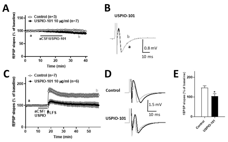
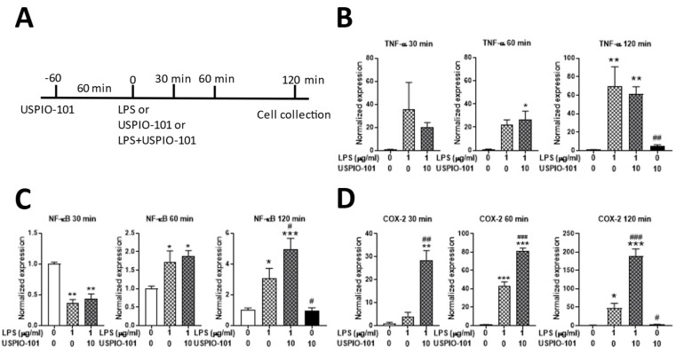
Our previous studies have revealed the ultrasmall superparamagnetic iron oxide in the amine group USPIO-101 has an analgesic effect on inflammatory pain. Here, we further investigated its effect on the spinal cord and brain via electrophysiological and molecular methods. We used a mouse inflammatory pain model, induced by complete Freund's adjuvant (CFA), and measured pain thresholds via von Frey methods. We also investigated the effects of USPIO-101 via an extracellular electrophysiological recording at the spinal dorsal horn synapses and hippocampal Schaffer collateral-CA1 synapses, respectively. The mRNA expression of pro-inflammatory cytokines was detected by quantitative real-time polymerase chain reaction (RT-qPCR). Our results showed intrathecal USPIO-101 produces similar analgesic behavior in mice with chronic inflammatory pain via intrathecal or intraplantar administration. The potentiated low-frequency stimulation-induced spinal cord long-term potentiation (LTP) at the spinal cord superficial dorsal horn synapses could decrease via USPIO-101 in mice with chronic inflammatory pain. However, the mRNA expression of cyclooxygenase-2 was enhanced with lipopolysaccharide (LPS) stimulation in microglial cells, and we also found USPIO-101 at 30 µg/mL could decrease the magnitude of hippocampal LTP. These findings revealed that intrathecal USPIO-101 presented an analgesia effect at the spinal cord level, but had neurotoxicity risk at higher doses.

Keywords: analgesia; inflammatory pain; long-term potentiation; neurotoxicity; pro-inflammatory cytokines; ultrasmall magnetic iron oxide nanoparticles.


Conflict of interest statement

The authors declare no conflict of interest.

Figures

Figure 1
Figure 1
The dynamic light scattering (DLS), zeta potential analysis, and Fourier transform infrared spectroscopy (FTIR) spectra of ultrasmall magnetic iron oxide nanoparticles. (A) number distribution of USPIO-101, as measured by DLS. The concentration of USPIO-101 was 1 mg/mL. (B) Zeta potential analysis of USPIO-101. (C) Fourier transform infrared (FTIR) spectra are presented for USPIO-101 (black) and USPIO-102 (red), respectively. The -NH2 and -CH2 group signals are expressed in USPIO-101 at the wavelength of 3300~3500 cm−1, 1560~1640 cm−1, or 1405~1465 cm−1 (arrow). The -COOH group signal is expressed in USPIO-102 at the wavelength of 1550~1610 cm−1 (arrow).
Figure 2
Figure 2
USPIO-101 attenuated chronic inflammatory pain via intrathecal or intraplantar injection. Mechanical pain sensitivity was measured using von Frey microfilaments. USPIO-101 attenuated the analgesia behavior in both (A) intrathecal and (B) intraplantar injection (10 mg/mL, 10 μL) after CFA paw injection for four days. The paw withdrawal thresholds were measured every 30 min until there was no difference between the three groups. Data were analyzed by two-way ANOVA and post-hoc with Tukey’s test. *: p < 0.05 vs. PBS sham group.
Figure 3
Figure 3
The effect of USPIO-101 on spinal cord LTP in CFA paw-injected mice. (A) Time courses of the slope of fEPSPs recorded before and after low-frequency stimulation (LFS, arrow) in spinal cord slices from CFA- or saline-treated mice. (C) The bar graph represents the magnitudes of potentiation, which averaged 20 fEPSPs recorded 30~40 min after LFS. **: p < 0.01 (unpaired t-tests) vs. CFA-treated ipsilateral group. (D) Time courses of the slope of fEPSPs recorded before and after LFS (arrow) in spinal cord slices from CFA-treated mice with USPIO-101 (10 or 30 µg/mL), respectively. USPIO-101 (10 or 30 µg/mL) applied for 7.5 min before LFS stimulation, respectively. (F) The bar graph represents the magnitudes of potentiation, which averaged 20 fEPSPs recorded 30~40 min after LFS at a concentration of 10 or 30 µg/mL. * or **: p < 0.05 or p < 0.01 vs. control group (one-way ANOVA). (B,E) Twenty recorded fEPSPs at time points a and b were averaged in each group. The slope of each fEPSP was expressed as % of the baseline fEPSP slope, which was the average of 20 fEPSPs at the beginning of 10 min recording. n indicates the number of slices recorded.
Figure 4
Figure 4
The effect of USPIO-101 on basal transmission (A) and spinal cord LTP (B,C) at the spinal cord superficial dorsal horn synapses, respectively. (A) After baseline recording for 10 min, USPIO-101 (10 µg/mL) was applied for another 15 min, then washed for 15 min. No difference was observed in the basal transmission after application of USPIO-101 to the spinal cord superficial dorsal horn synapses. (C) Time courses of the slope of fEPSPs recorded before and after LFS (arrow) in spinal cord slices. Drugs: USPIO-101 (10 µg/mL) was applied for 7.5 min before LFS stimulation. (E) The bar graph represents the magnitudes of potentiation, which averaged 20 fEPSPs recorded 30~40 min after LFS. *: p < 0.05 vs. control group (one-way ANOVA). (B,D) The traces shown in the graph are averaged 20 recordings of fEPSPs at times a and b in each group. The expression and analysis of the baseline fEPSP slope are the same as Figure 2. n indicates the number of slices recorded.
Figure 5
Figure 5
Intracellular reactive oxygen species level after treatment with USPIO-101 in SH-SY5Y or SM826 cells. (A,B) Hydrogen peroxide (H2O2) as positive control and USPIO-101 (10 or 30 μg/mL) were used in SH-SY5Y cells for 30 min (A, H2O2 1 mM) and 24 h (B, H2O2 0.2 mM), respectively. (C) Lipopolysaccharide (LPS) 1 μg/mL as positive control and USPIO-101 (10 or 30 μg/mL) were used in SM826 cells for 24 h, respectively. RFU means the relative fluorescence unit in 3 × 10⁴ cells/well in 96-well plates. Data shown are the mean ± SEM of three independent experiments performed in triplicate. * or ***: p < 0.05 or p < 0.001 vs. control group. ###: p < 0.001 vs. the H2O2 group (one-way ANOVA).
Figure 6
Figure 6
USPIO-101 potentiates the LPS-induced NF-kB and COX-2 mRNA expression in SM826 microglial cells. (A) The time scale for treating USPIO-101 and LPS stimulation. (BD) The levels of mRNAs encoding the pro-inflammatory cytokines TNF-α (B), NF-κB (C), and COX-2 (D) were analyzed by qRT-PCR and normalized by the expression of GAPDH (glyceraldehyde-3-phosphate dehydrogenase) at three time points after LPS stimulation: 30, 60, or 120 min. Data are presented as mean ± SEM (n = 4). * or ** or ***: p < 0.05, or p < 0.01, or p < 0.001 vs. control; # or ## or ###: p < 0.05, or p < 0.01, or p < 0.001 vs. LPS.
Figure 7
Figure 7
The effect of USPIO-101 on hippocampal LTP. (A) Time courses of the slope of fEPSPs recorded before and after theta-burst stimulation (TBS, arrow) in hippocampal slices. Drugs: USPIO-101 (10 or 30 µg/mL) was applied for 7.5 min before TBS stimulation. (B) The traces shown in the graph are the average of 20 recorded fEPSPs at times a and b in each group. (C) The bar graph represents the magnitudes of potentiation, with the average of 20 fEPSPs recorded 30~40 min after TBS at a concentration of 10 or 30 µg/mL. *: p < 0.05 vs. control group. #: p < 0.05 vs. USPIO-101 group (one-way ANOVA). n indicates the number of slices recorded. The expression and analysis of the baseline fEPSP slope are the same as Figure 3.

References

    1. Alphandery E. Iron oxide nanoparticles for therapeutic applications. Drug Discov. Today. 2020;25:141–149. doi: 10.1016/j.drudis.2019.09.020. - DOI - PubMed
    1. Dadfar S.M., Roemhild K., Drude N.I., von Stillfried S., Knuchel R., Kiessling F., Lammers T. Iron oxide nanoparticles: Diagnostic, therapeutic and theranostic applications. Adv. Drug Deliv. Rev. 2019;138:302–325. doi: 10.1016/j.addr.2019.01.005. - DOI - PMC - PubMed
    1. Andreu V., Arruebo M. Current progress and challenges of nanoparticle-based therapeutics in pain management. J. Control. Release Off. J. Control. Release Soc. 2018;269:189–213. doi: 10.1016/j.jconrel.2017.11.018. - DOI - PubMed
    1. Chen J., Jin T., Zhang H. Nanotechnology in Chronic Pain Relief. Front. Bioeng. Biotechnol. 2020;8:682. doi: 10.3389/fbioe.2020.00682. - DOI - PMC - PubMed
    1. Wu P.C., Hsiao H.T., Lin Y.C., Shieh D.B., Liu Y.C. The analgesia efficiency of ultrasmall magnetic iron oxide nanoparticles in mice chronic inflammatory pain model. Nanomed. Nanotechnol. Biol. Med. 2017;13:1975–1981. doi: 10.1016/j.nano.2017.05.005. - DOI - PubMed