Review
. 2022 Feb 17;14(2):434.
doi: 10.3390/pharmaceutics14020434.

Recent Development of Drug Delivery Systems through Microfluidics: From Synthesis to Evaluation

Affiliations
Review

Recent Development of Drug Delivery Systems through Microfluidics: From Synthesis to Evaluation

Zhiyuan Ma et al. Pharmaceutics. .

Abstract

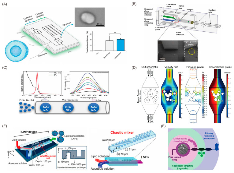
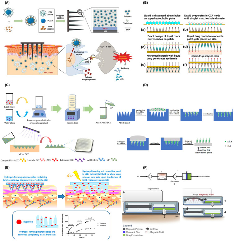
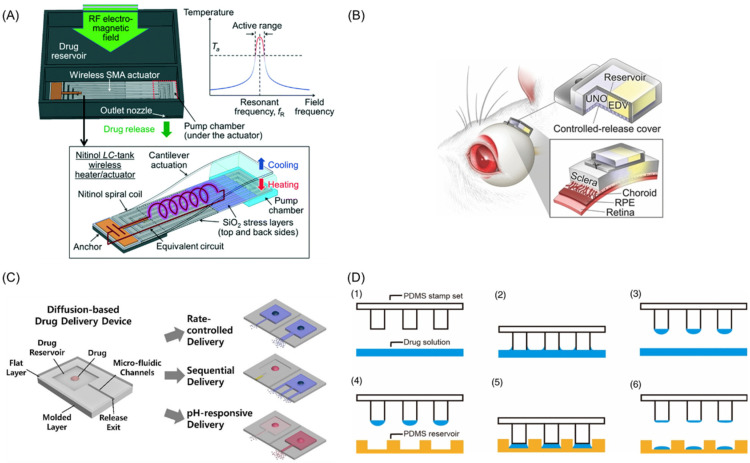
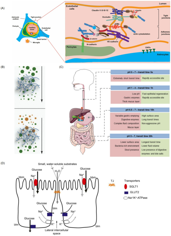
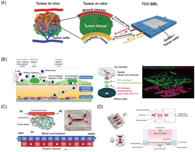
Conventional drug administration usually faces the problems of degradation and rapid excretion when crossing many biological barriers, leading to only a small amount of drugs arriving at pathological sites. Therapeutic drugs delivered by drug delivery systems to the target sites in a controlled manner greatly enhance drug efficacy, bioavailability, and pharmacokinetics with minimal side effects. Due to the distinct advantages of microfluidic techniques, microfluidic setups provide a powerful tool for controlled synthesis of drug delivery systems, precisely controlled drug release, and real-time observation of drug delivery to the desired location at the desired rate. In this review, we present an overview of recent advances in the preparation of nano drug delivery systems and carrier-free drug delivery microfluidic systems, as well as the construction of in vitro models on-a-chip for drug efficiency evaluation of drug delivery systems. We firstly introduce the synthesis of nano drug delivery systems, including liposomes, polymers, and inorganic compounds, followed by detailed descriptions of the carrier-free drug delivery system, including micro-reservoir and microneedle drug delivery systems. Finally, we discuss in vitro models developed on microfluidic devices for the evaluation of drug delivery systems, such as the blood-brain barrier model, vascular model, small intestine model, and so on. The opportunities and challenges of the applications of microfluidic platforms in drug delivery systems, as well as their clinical applications, are also discussed.

Keywords: carrier-free; drug delivery system; in vitro model; micro-reservoir; microfluidic; microneedles.


Conflict of interest statement

The authors declare no conflict of interest.

Figures

Figure 1
Figure 1
Different approaches for the fabrication of nanoparticles. (A) Schematic of the microfluidic device for one-step formation of plasmid DNA (pDNA)/cationic liposome (CL) lipoplexes and its in vitro efficacy. (B) Schematic of the flow-focusing device used to engineer and monitor Janus drug particle formation. (C) Schematic of the microfluidic reactor used for the synthesis of ZnSe quantum dots. (D) The simulated microfluidic mixer unit with two inlets and two outlets. (E) Three-dimensional views and top views of the iLiNP device. (F) The strategy employed for dual-targeted two-drug nanocarriers based on mesoporous silica nanoparticles. (A) Balbino et al. [27]; (B) Sundararajan et al. [31]; (C) Guidelli et al. [36]; (D) Wang et al. [24]; (E) Kimura et al. [21]; (F) Castillo et al. [38].
Figure 2
Figure 2
(A) Working principle diagram of ph1n1 DNA vaccine delivery platform of PLGA/PEI nanoparticle MNs. (B) Schematic diagram of MNs medical coating system. (a) liquid is dispensed above holes, (b) liquid evaporates in CCA mode until droplet matches hole diameter, (c) exact dosage of liquid coats MNs on patch, (d) liquid drug coated MN patch gets placed on skin, (e) MN patch with liquid drug penetrates epidermis and (f) liquid drug stays in skin. (C) Process diagram of the ACO–NLCs–MNs synthesis. (D) The process of fabricating ALA–HA fast-dissolving MNs. (E) Working principle of the light-responsive hydrogel MNs. (F) Process flow diagram of the magnetic polymer-driven MNs system. (A) HaeYong et al. [64]; (B) Andreas et al. [65]; (C) Guo et al. [67]; (D) Zhao et al. [68]; (E) John G. et al. [69]; (F) Jayaneththi et al. [74].
Figure 3
Figure 3
(A) Schematic diagram of wireless implantable magnetic driven DDSs and its working principle. (B) Working diagram of dual-drug delivery system. (C) The “reservoir-microfluidic channel” system optimization and drug release diagram. (D) Schematic diagram of the wet μCP system manufacturing: (1) PDMS stamp set and drug solution, (2) tip contact with drug solution, (3) drug coated stamps, (4) The position of drug coated stamp above PDMS reservoir, (5) stamp contact on a target surface and (6) final drug loading formation. (A) Jeffrey Fong et al. [80]; (B) Nobuhiro et al. [82]; (C) Yang et al. [84]; (D) Lee et al. [85].
Figure 4
Figure 4
(A) Cells association at and molecular organization of the neurovascular unit (NVU). (B) The steric (up) and interactive (down) barrier properties of mucus. The mucus forms a filter, which can prevent diffusion across the gel according to size (up) or surface properties (down) of the diffusing compound. (C) Schematic representation of GIT sections and their pH, transit time, and relevant parameters for drug delivery. (D) Schematic diagram illustrating glucose absorption by transcellular pathways and paracellular pathways. Reprinted and adapted with permission from: (A) Kadry et al. [89], (B) Lieleg et al. [108], (C) Alonso et al. [129], and (D) Karasov et al. [130].
Figure 5
Figure 5
Different kinds of organs-on-a-chip. (A) The biomimetic in vitro model of tumor-on-a-chip with bio-printed blood and lymphatic vessel pair. (B) Multilayered blood vessel/tumor tissue chip (MBTC). (C) The schematic for the system of tumor tissue and blood vessels (a) schematic (b) picture of microfluidic device. (c) Computational mesh (D) The assembled microfluidic human model of BBB and its side view showing the fluid pathway. Reprinted and adapted with permission from: (A) Cao et al. [101], (B) Lee et al. [114], (C) Li et al. [100], and (D) Wang et al. [121].

References

    1. Li C., Wang J., Wang Y., Gao H., Wei G., Huang Y., Yu H., Gan Y., Wang Y., Mei L., et al. Recent progress in drug delivery. Acta Pharm. Sin. B. 2019;9:1145–1162. doi: 10.1016/j.apsb.2019.08.003. - DOI - PMC - PubMed
    1. Sanjay S.T., Zhou W., Dou M., Tavakoli H., Ma L., Xu F., Li X. Recent advances of controlled drug delivery using microfluidic platforms. Adv. Drug Deliv. Rev. 2018;128:3–28. doi: 10.1016/j.addr.2017.09.013. - DOI - PMC - PubMed
    1. Park K. Controlled drug delivery systems: Past forward and future back. J. Control. Release. 2014;190:3–8. doi: 10.1016/j.jconrel.2014.03.054. - DOI - PMC - PubMed
    1. Cochrane A., Albers H.J., Passier R., Mummery C.L., van den Berg A., Orlova V.V., van der Meer A.D. Advanced in vitro models of vascular biology: Human induced pluripotent stem cells and organ-on-chip technology. Adv. Drug Deliv. Rev. 2019;140:68–77. doi: 10.1016/j.addr.2018.06.007. - DOI - PubMed
    1. Bendre A., Bhat M.P., Lee K.-H., Altalhi T., Alruqi M.A., Kurkuri M. Recent developments in microfluidic technology for synthesis and toxicity-efficiency studies of biomedical nanomaterials. Mater. Today Adv. 2022;13:100205. doi: 10.1016/j.mtadv.2022.100205. - DOI

LinkOut - more resources