. 2022 Feb 19;22(4):1629.
doi: 10.3390/s22041629.

Multi-Method Diagnosis of Blood Microscopic Sample for Early Detection of Acute Lymphoblastic Leukemia Based on Deep Learning and Hybrid Techniques

Affiliations

Multi-Method Diagnosis of Blood Microscopic Sample for Early Detection of Acute Lymphoblastic Leukemia Based on Deep Learning and Hybrid Techniques

Ibrahim Abunadi et al. Sensors (Basel). .

Abstract

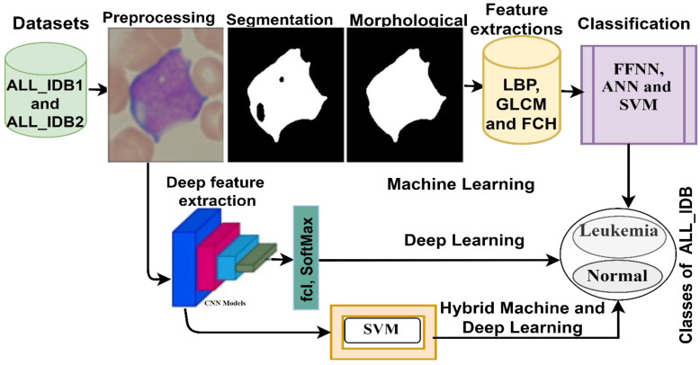
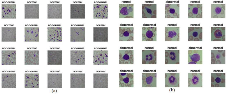
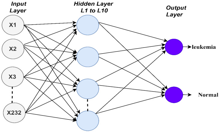
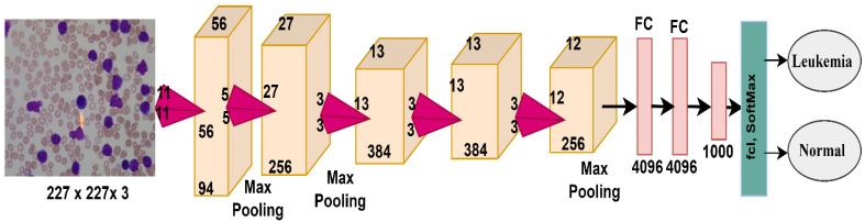
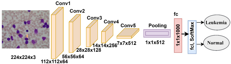
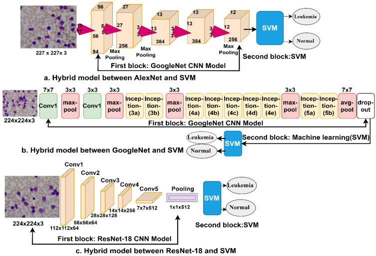
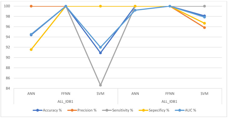
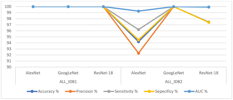
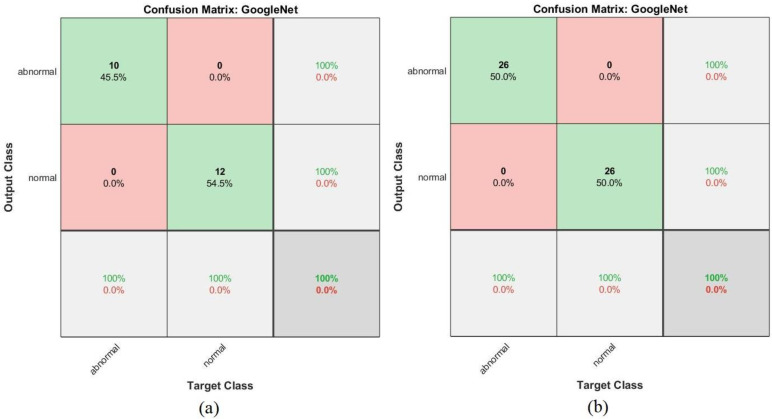
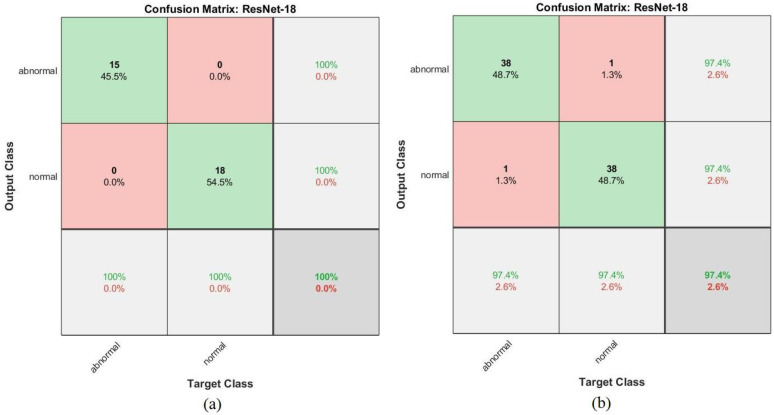
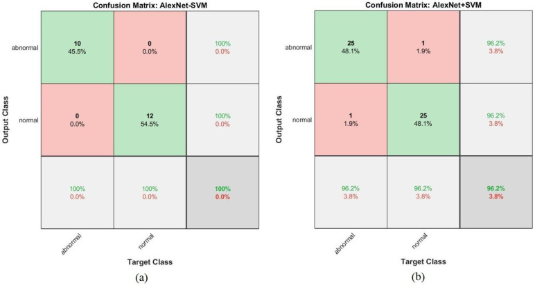
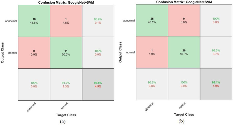
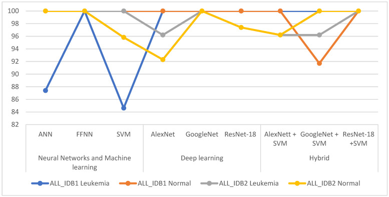
Leukemia is one of the most dangerous types of malignancies affecting the bone marrow or blood in all age groups, both in children and adults. The most dangerous and deadly type of leukemia is acute lymphoblastic leukemia (ALL). It is diagnosed by hematologists and experts in blood and bone marrow samples using a high-quality microscope with a magnifying lens. Manual diagnosis, however, is considered slow and is limited by the differing opinions of experts and other factors. Thus, this work aimed to develop diagnostic systems for two Acute Lymphoblastic Leukemia Image Databases (ALL_IDB1 and ALL_IDB2) for the early detection of leukemia. All images were optimized before being introduced to the systems by two overlapping filters: the average and Laplacian filters. This study consists of three proposed systems as follows: the first consists of the artificial neural network (ANN), feed forward neural network (FFNN), and support vector machine (SVM), all of which are based on hybrid features extracted using Local Binary Pattern (LBP), Gray Level Co-occurrence Matrix (GLCM) and Fuzzy Color Histogram (FCH) methods. Both ANN and FFNN reached an accuracy of 100%, while SVM reached an accuracy of 98.11%. The second proposed system consists of the convolutional neural network (CNN) models: AlexNet, GoogleNet, and ResNet-18, based on the transfer learning method, in which deep feature maps were extracted and classified with high accuracy. All the models obtained promising results for the early detection of leukemia in both datasets, with an accuracy of 100% for the AlexNet, GoogleNet, and ResNet-18 models. The third proposed system consists of hybrid CNN-SVM technologies, consisting of two blocks: CNN models for extracting feature maps and the SVM algorithm for classifying feature maps. All the hybrid systems achieved promising results, with AlexNet + SVM achieving 100% accuracy, Goog-LeNet + SVM achieving 98.1% accuracy, and ResNet-18 + SVM achieving 100% accuracy.

Keywords: acute lymphoblastic leukemia; convolutional neural network; fuzzy color histogram; gray level co-occurrence matrix; hybrid method; local binary pattern; machine learning.


Conflict of interest statement

The authors declare no conflict of interest.

Figures

Figure 1
Figure 1
Morphological diagram for leukemia diagnosis from the ALL_IDB1 and ALL_IDB2 dataset.
Figure 2
Figure 2
Samples from the (a) ALL-IDB1 and (b) ALL-IDB2 datasets.
Figure 3
Figure 3
Samples from the (a) ALL-IDB1 and (b) ALL-IDB2 datasets after enhancement.
Figure 4
Figure 4
Samples from the two datasets after segmentation.
Figure 5
Figure 5
Samples from the two datasets before and after the morphological processes.
Figure 6
Figure 6
Fusion of features extracted by the LBP, GLCM, and FCH algorithms.
Figure 7
Figure 7
Architecture of the artificial neural network and feed forward neural network classifiers of the ALL-IDB dataset.
Figure 8
Figure 8
AlexNet network structure for ALL-IDB dataset diagnostics.
Figure 9
Figure 9
GoogLeNet network structure for ALL-IDB dataset diagnostics.
Figure 10
Figure 10
ResNet-18 network structure for ALL-IDB dataset diagnostics.
Figure 11
Figure 11
Deep learning–machine learning hybrid techniques: (a) AlexNet + SVM; (b) GoogleNet + SVM; and (c) ResNet-18 + SVM.
Figure 12
Figure 12
Training of the feed forward neural network classifier of the ALL_IDB1 and ALL_IDB2 datasets.
Figure 13
Figure 13
Performance of the artificial neural network classifier of the (a) ALL_IDB1 and (b) ALL_IDB2 datasets.
Figure 14
Figure 14
Gradient and validation check values of the (a) ALL_IDB1 and (b) ALL_IDB2 datasets.
Figure 15
Figure 15
Receiver operating characteristic values of the (a) ALL_IDB1 and (b) ALL_IDB2 datasets.
Figure 16
Figure 16
Error histograms of the (a) ALL_IDB1 and (b) ALL_IDB2 datasets.
Figure 17
Figure 17
Regression of the (a) ALL_IDB1 and (b) ALL_IDB2 datasets.
Figure 18
Figure 18
Confusion matrix for the ANN algorithm of the (a) ALL_IDB1 and (b) ALL_IDB2 datasets.
Figure 19
Figure 19
Confusion matrix for the FFNN algorithm of the (a) ALL_IDB1 and (b) ALL_IDB2 datasets.
Figure 20
Figure 20
Performances of all the ANN, FFNN and SVM algorithms for the ALL_IDB1 and ALL_IDB2 datasets.
Figure 21
Figure 21
Performances of all the convolutional neural network models for the ALL_IDB1 and ALL_IDB2 datasets.
Figure 22
Figure 22
Confusion matrix for the AlexNet model for the (a) ALL_IDB1 and (b) ALL_IDB2 datasets.
Figure 23
Figure 23
Confusion matrix for the GoogLeNet model for the (a) ALL_IDB1 and (b) ALL_IDB2 datasets.
Figure 24
Figure 24
Confusion matrix for the ResNet-18 model for the (a) ALL_IDB1 and (b) ALL_IDB2 datasets.
Figure 25
Figure 25
Performances of all the hybrid techniques for the ALL_IDB1 and ALL_IDB2 datasets.
Figure 26
Figure 26
Confusion matrix for the AlexNet + SVM hybrid technique for the (a) ALL_IDB1 and (b) ALL_IDB2 datasets.
Figure 27
Figure 27
Confusion matrix for the GoogLeNet + SVM hybrid technique for the (a) ALL_IDB1 and (b) ALL_IDB2 datasets.
Figure 28
Figure 28
Confusion matrix for the ResNet-18 + SVM hybrid technique for the (a) ALL_IDB1 and (b) ALL_IDB2 datasets.
Figure 29
Figure 29
Comparison of the performances of the proposed systems for diagnosing leukemia from the two datasets.
Figure 30
Figure 30
Comparing the performance of our proposed system with the previous systems.

Similar articles

Cited by

References

    1. Kuan D.H., Wu C.C., Su W.Y., Huang N.T. A microfluidic device for simultaneous extraction of plasma, red blood cells, and on-chip white blood cell trapping. Sci. Rep. 2018;8:1–9. doi: 10.1038/s41598-018-33738-8. - DOI - PMC - PubMed
    1. Farag M.R., Alagawany M. Erythrocytes as a biological model for screening of xenobiotics toxicity. Chem. Biol. Interact. 2018;279:73–83. doi: 10.1016/j.cbi.2017.11.007. - DOI - PubMed
    1. Rezatofighi S.H., Soltanian-Zadeh H. Automatic recognition of five types of white blood cells in peripheral blood. Comput. Med. Imaging Graph. 2011;35:333–343. doi: 10.1016/j.compmedimag.2011.01.003. - DOI - PubMed
    1. Faggio C., Sureda A., Morabito S., Sanches-Silva A., Mocan A., Nabavi S.F., Nabavi S.M. Flavonoids and platelet aggregation: A brief review. Eur. J. Pharmacol. 2017;807:91–101. doi: 10.1016/j.ejphar.2017.04.009. - DOI - PubMed
    1. Al-Hafiz F., Al-Megren S., Kurdi H. Red blood cell segmentation by thresholding and Canny detector. Procedia Comput. Sci. 2018;141:327–334. doi: 10.1016/j.procs.2018.10.193. - DOI

LinkOut - more resources