. 2022 Jan 22;10(2):173.
doi: 10.3390/vaccines10020173.

Immunization with GP1 but Not Core-like Particles Displaying Isolated Receptor-Binding Epitopes Elicits Virus-Neutralizing Antibodies against Junín Virus

Affiliations

Immunization with GP1 but Not Core-like Particles Displaying Isolated Receptor-Binding Epitopes Elicits Virus-Neutralizing Antibodies against Junín Virus

Gleyder Roman-Sosa et al. Vaccines (Basel). .

Abstract

New World arenaviruses are rodent-transmitted viruses and include a number of pathogens that are responsible for causing severe human disease. This includes Junín virus (JUNV), which is the causative agent of Argentine hemorrhagic fever. The wild nature and mobility of the rodent reservoir host makes it difficult to control the disease, and currently passive immunization with high-titer neutralizing antibody-containing plasma from convalescent patients is the only specific therapy. However, dwindling supplies of naturally available convalescent plasma, and challenges in developing similar resources for other closely related viruses, have made the development of alternative antibody-based therapeutic approaches of critical importance. In this study, we sought to induce a neutralizing antibody response in rabbits against the receptor-binding subunit of the viral glycoprotein, GP1, and the specific peptide sequences in GP1 involved in cellular receptor contacts. While these specific receptor-interacting peptides did not efficiently induce the production of neutralizing antibodies when delivered as a particulate antigen (as part of hepatitis B virus core-like particles), we showed that recombinant JUNV GP1 purified from transfected mammalian cells induced virus-neutralizing antibodies at high titers in rabbits. Further, neutralization was observed across a range of unrelated JUNV strains, a feature that is critical for effectiveness in the field. These results underscore the potential of GP1 alone to induce a potent neutralizing antibody response and highlight the importance of epitope presentation. In addition, effective virus neutralization by rabbit antibodies supports the potential applicability of this species for the future development of immunotherapeutics (e.g., based on humanized monoclonal antibodies). Such information can be applied in the design of vaccines and immunogens for both prevention and specific therapies against this and likely also other closely related pathogenic New World arenaviruses.

Keywords: GP1; Junín virus; arenavirus; glycoprotein; immune response; neutralizing antibodies.


Conflict of interest statement

The authors declare no conflict of interest. The sponsors had no role in the design, execution, interpretation, or writing of the study.

Figures
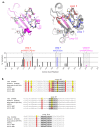
Figure 1
Figure 1
Strategy for the generation of Junín virus (JUNV)-derived peptide antigens for expression on hepatitis B virus core-like particles. (A) Crystal structures of Machupo virus (MACV) GP1 and JUNV GP1. The X-ray structure of MACV GP1 in complex with transferrin receptor 1 (TfR1) (PDB:3KAS [11]), in which the TfR1 atoms were removed, is represented in the left panel. GP1 is colored pink except for the residues making contact with TfR1 in the complex, which are shown as sticks (color coded according to the atom type: carbon, light grey; nitrogen, blue; oxygen, red; sulfur, yellow). The right panel shows an overlay of the same structure with JUNV GP1 (extracted from the X-ray structure of JUNV GP1 in complex with Fab GD01, PDB:5EN2 [12]). The MACV GP1 structure is represented with its interacting residues as sticks, as in the left panel, except that the rest of the protein is in grey. The JUNV GP1 structure is shown only as ribbons in which the three loops tested are colored, as indicated. Note that Loop 3 extends all along the interaction surface and could constitute a potential linear epitope eliciting neutralizing antibodies. These two panels were prepared with the PyMOL Molecular Graphics System (Version 2.1, Schrödinger). Conservation of the amino acids in and around the respective loops (amino acid positions 80–240) are shown based on an alignment of the sequences of 63 unique naturally occurring JUNV isolates available in GenBank (Figure S2). The positions of the respective loops are boxed and bars indicating the frequency of variable amino acids within these regions are shown in color. The corresponding peptide sequences are shown, and asterisks indicate these variable sites. (B) Sequence alignment for JUNV strains used in this study. Details of the sequence alignment are shown for the region from amino acid 80 to 240 for the CbaIV4454 strain (GenBank: DQ272266), against which the peptide sequences were designed, and the other strains used for the testing of cross-neutralization in this study. Sequences corresponding to the selected peptide loops are indicated in bold text and underlined. Sequences for strains Romero (GenBank: JN801476), Espindola (GenBank: DQ854739), and P3551 (GenBank: MZ408913, this study), and both a reference sequence for strain XJ13 (XJ13-REF; GenBank: FJ805378) and the sequence determined based on the XJ13 isolate available in our laboratory (XJ13-EXP; GenBank: MZ408914) are shown. Sites showing amino acid variation are highlighted in color, with the amino acid observed in strain CbaIV4454 shown in yellow and all other variants shown in red. *: “asterisks indicate these variable sites“.
Figure 2
Figure 2
Preparation of antigens for immunization. (A) Purification of recombinant Junín virus (JUNV) GP1. JUNV GP1 containing a C-terminal Strep tag (GP1-Strep) was transiently expressed in HEK-293 cells and purified via affinity chromatography using Strep-Tactin resin. The eluted fractions were analyzed by Coomassie staining (left panel). Alternatively, JUNV GP1 containing a C-terminal hexahistidine (GP1-His) tag was expressed in Freestyle 293-F cells and purified via affinity chromatography using HisTrap columns. The resulting protein fraction was also analyzed by Coomassie staining (right panel). (B) Hepatitis B virus core-like particles (HBV-CLPs) carrying JUNV GP1 peptide loops. HBV-CLPs containing the indicated antigens (i.e., Loop 3, Loop 7, Loop 10, or a control FLAG tag) were produced in E. coli and isolated from the soluble fraction of lysates via Strep tag-mediated affinity purification. Eluates were analyzed by Coomassie staining (upper panel) and Western blot with an anti-Strep antibody (1:30,000; StrepMAB Classic-HRP; IBA Lifesciences, Göttingen, Germany; lower panel). The position of the HBV core protein is indicated. (C) Electron microscopic analysis of HBV-CLPs. The purified HBV-CLPs generated in (B) were further analyzed by electron microscopy with negative staining. The scale bar shown represents 80 nm.
Figure 3
Figure 3
Development of antibodies following vaccination with recombinant Junín virus (JUNV) GP1. Blood samples were collected from rabbits either before immunization or 14 days after each of 3 immunizations with 80 ug of recombinant JUNV GP1-Strep. A sample from another rabbit unrelated to this study served as an additional control (Neg Ctrl). Serum fractions were diluted 1:10 for use in an indirect ELISA assay with JUNV GP1-Strep, JUNV-GP1-His, or Schmallenberg virus Gc amino-Strep (SBV-Gc-Strep) as the target antigen, as indicated. Data from at least two independent experiments are represented and the results of a one-way ANOVA comparing samples post-vaccination to the respective pre-immunization samples are shown (ns not significant; * p ≤ 0.05; *** p ≤ 0.001; **** p ≤ 0.0001).
Figure 4
Figure 4
Assessment of neutralizing antibody development following vaccination with recombinant Junín virus (JUNV) GP1 based on neutralization of transcription and replication-competent virus-like particles (trVLPs). (A) Development of neutralizing antibodies in response to vaccination. trVLPs containing the GP of JUNV (strain Romero) were incubated 1:1 (i.e., final 1:2 dilution) with serum from rabbits vaccinated with rGP1-Strep collected before or during the immunization process, as indicated. Serum from human Candid#1 vaccinees (Vacc Ctrl) or a control pooled human AB serum served as additional controls. Samples were incubated for 2 h at 37 °C before being used to infect Huh7 cells pre-transfected with plasmids expressing JUNV NP and L. After 48 h, these cells were harvested and their nanoluciferase activity measured (as a measure of viral RNA synthesis). Data from at least two independent experiments are represented and the results of a one-way ANOVA comparison to control antibody treated samples are shown. (B) Titration of neutralizing antibodies. Neutralization of trVLPs was performed as in (A) using the serum collected after the final immunization. The sera were prepared as a 1:2 dilution series before being added to trVLPs to achieve a dilution range from 1:2 to 1:512. Data from at least three independent experiments are represented and are shown relative to values obtained with the negative control sera. The results of a one-way ANOVA comparing samples to the negative control serum are shown. (C) Neutralization of trVLPs expressing JUNV GP from different virus strains. JUNV trVLPs expressing heterologous GP proteins from four different JUNV strains (i.e., Romero, Espindola, P3551, and XJ13) were generated and used in neutralization assays as described in (A) with the serum samples collected after the final immunization. Data from at least two independent experiments are represented and the results of a one-way ANOVA to compare samples to a negative control serum are shown (ns not significant; * p ≤ 0.05; ** p ≤ 0.01; *** p ≤ 0.001; **** p ≤ 0.0001).
Figure 5
Figure 5
Assessment of neutralizing antibody development following vaccination with recombinant Junín virus (JUNV) GP1 based on the plaque reduction neutralization test. JUNV (strain Cba IV4454) diluted to 103 pfu/mL was mixed 1:1 with serum from rabbits vaccinated with recombinant JUNV GP1-Strep or from a naïve rabbit. Serum from human Candid#1 vaccinees (Vacc Ctrl) or a control pooled human AB serum served as additional controls. Samples were incubated for 1 h at 37 °C before 100 μL were used to infect Vero cells for analysis using plaque assay. Values are expressed as a percentage of the number of plaques in samples treated with the control serum (i.e., naïve rabbit or human AB serum). Data from at least two independent experiments are represented and the results of a one-way ANOVA comparison of experimental samples with the control antibody are shown (ns not significant; * p ≤ 0.05; ** p ≤ 0.01; *** p ≤ 0.001; **** p ≤ 0.0001).
Figure 6
Figure 6
Development of antibodies following vaccination with Hepatitis B virus core-like particles (HBV-CLPs) expressing Junín virus (JUNV)-derived peptides. Blood samples were collected from rabbits either before immunization or 14 days after each of 3 immunizations with 80 µg of purified HBV-CLPs expressing 1 of 3 JUNV-derived peptides (i.e., Loop 3, Loop 7, Loop 10), as indicated. Vaccination with HBV-CLPs without a JUNV-specific antigen (i.e., HBV-CLP (FLAG)) and a sample from another rabbit unrelated to this study (Neg Ctrl) served as additional controls. Serum fractions isolated from these samples were diluted 1:10 for use in an indirect ELISA assay with JUNV GP1-Strep, JUNV-GP1-His, or Schmallenberg virus Gc amino-Strep (SBV-Gc-Strep) as the target antigen, as indicated. Data from at least two independent experiments are represented and the results of a one-way ANOVA comparing post-vaccination samples to the respective pre-bleed samples are shown (ns not significant; * p ≤ 0.05; ** p ≤ 0.01; *** p ≤ 0.001; **** p ≤ 0.0001).
Figure 7
Figure 7
Assessment of neutralizing antibody development following vaccination with Hepatitis B virus core-like particles (HBV-CLPs) expressing Junín virus (JUNV)-derived peptides using neutralization of transcription and replication-competent virus-like particles (trVLPs). trVLPs containing the GP of JUNV (strain Romero) were incubated 1:1 (i.e., final 1:2 dilution) with serum from rabbits vaccinated with HBV-CLPs expressing 1 of 3 JUNV-derived peptides (i.e., Loop 3, Loop 7, Loop 10) collected before or during the immunization process, as indicated. Serum from human Candid#1 vaccinees (Vacc Ctrl) or a control pooled human AB serum served as additional controls. Samples were incubated for 2 h at 37 °C before being used to infect Huh7 cells pre-transfected with plasmids expressing JUNV NP and L. After 48 h, these cells were harvested and their nanoluciferase activity measured (as a measure of viral RNA synthesis). Data from at least two independent experiments are represented and the results of a one-way ANOVA comparison with control antibody-treated samples are shown. (B) Titration of neutralizing antibodies. Neutralization of trVLPs was performed as in (A) using the serum collected after the final immunization. The sera were prepared as a 1:2 dilution series before being added to trVLPs to achieve a dilution range from 1:2 to 1:128. Data from four independent experiments are represented and are shown relative to values obtained with the negative control sera (i.e., HBV-CLP-FLAG or human AB serum). The results of a one-way ANOVA comparing samples to the negative control serum are shown (ns not significant; * p ≤ 0.05; ** p ≤ 0.01; *** p ≤ 0.001; **** p ≤ 0.0001).
Figure 8
Figure 8
Assessment of neutralizing antibody development following vaccination with hepatitis B virus core-like particles (HBV-CLPs) expressing Junín virus (JUNV)-derived peptides based on the plaque reduction neutralization test. JUNV (strain Cba IV4454) diluted to 103 pfu/mL was mixed 1:1 with serum from rabbits vaccinated with 1 of the HBV-CLPs indicated (Loop 3, Loop 7, or Loop 10), a pooled mixture of the sera from these rabbits, or serum from a rabbit that received a control HBV-CLP containing a FLAG peptide. Serum from human Candid#1 vaccinees (Vacc Ctrl) or a control pooled human AB serum served as additional controls. Samples were incubated for 1 h at 37 °C before 100 μL were used to infect Vero cells for analysis using the plaque assay. Values are expressed as a percentage of the number of plaques in samples treated with the control serum (i.e., HBV-CLP-FLAG or human AB serum). Data from at least two independent experiments are represented and the results of a one-way ANOVA comparison of the experimental samples with the control antibody are shown (ns not significant; * p ≤ 0.05; ** p ≤ 0.01; *** p ≤ 0.001).

Similar articles

Cited by

References

    1. Monath T.P. A short history of Lassa fever: The first 10–15 years after discovery. Curr. Opin. Virol. 2019;37:77–83. doi: 10.1016/j.coviro.2019.06.005. - DOI - PubMed
    1. Sarute N., Ross S.R. New World Arenavirus Biology. Annu. Rev. Virol. 2017;4:141–158. doi: 10.1146/annurev-virology-101416-042001. - DOI - PMC - PubMed
    1. Peters C.J. Human infection with arenaviruses in the Americas. Curr. Top. Microbiol. Immunol. 2002;262:65–74. doi: 10.1007/978-3-642-56029-3_3. - DOI - PubMed
    1. Tesh R.B. Viral hemorrhagic fevers of South America. Biomedica. 2002;22:287–295. doi: 10.7705/biomedica.v22i3.1166. - DOI - PubMed
    1. Mills J.N., Ellis B.A., Childs J.E., McKee K.T., Maiztegui J.I., Peters C.J., Ksiazek T.G., Jahrling P.B. Prevalence of infection with Junin virus in rodent populations in the epidemic area of Argentine hemorrhagic fever. Am. J. Trop. Med. Hyg. 1994;51:554–562. doi: 10.4269/ajtmh.1994.51.554. - DOI - PubMed

LinkOut - more resources