. 2022 Feb 10;10(2):271.
doi: 10.3390/vaccines10020271.

A Lateral Flow Immunoassay Coupled with a Spectrum-Based Reader for SARS-CoV-2 Neutralizing Antibody Detection

Affiliations

A Lateral Flow Immunoassay Coupled with a Spectrum-Based Reader for SARS-CoV-2 Neutralizing Antibody Detection

Rui-Lin Huang et al. Vaccines (Basel). .

Abstract

As of August 2021, there have been over 200 million confirmed case of coronavirus disease 2019 caused by severe acute respiratory syndrome coronavirus and more than 4 million COVID-19-related deaths globally. Although real-time polymerase chain reaction is considered to be the primary method of detection for SARS-CoV-2 infection, the use of serological assays for detecting COVID-19 antibodies has been shown to be effective in aiding with diagnosis, particularly in patients who have recovered from the disease and those in later stages of infection. Since it has a high detection rate and few limitations compared to conventional enzyme-linked immunosorbent assay protocols, we used a lateral flow immunoassay as our diagnostic tool of choice. Since lateral flow immunoassay results interpreted by the naked eye may lead to erroneous diagnoses, we developed an innovative, portable device with the capacity to capture a high-resolution reflectance spectrum as a means of promoting diagnostic accuracy. We combined this spectrum-based device with commercial lateral flow immunoassays to detect the neutralizing antibody in serum samples collected from 30 COVID-19-infected patients (26 mild cases and four severe cases). The results of our approach, lateral flow immunoassays coupled with a spectrum-based reader, demonstrated a 0.989 area under the ROC curve, 100% sensitivity, 95.7% positive predictive value, 87.5% specificity, and 100% negative predictive value. As a result, our approach exhibited great value for neutralizing antibody detection. In addition to the above tests, we also tested plasma samples from 16 AstraZeneca-vaccinated (ChAdOx1nCoV-19) patients and compared our approach and enzyme-linked immunosorbent assay results to see whether our approach could be applied to vaccinated patients. The results showed a high correlation between these two approaches, indicating that the lateral flow immunoassay coupled with a spectrum-based reader is a feasible approach for diagnosing the presence of a neutralizing antibody in both COVID-19-infected and vaccinated patients.

Keywords: AstraZeneca; coronavirus disease 2019 (COVID-19); lateral flow immunoassay; neutralizing antibody; severe acute respiratory syndrome coronavirus-2 (SARS-CoV-2).


Conflict of interest statement

The authors declare no conflict of interest.

Figures

Figure 1
Figure 1
SARS-CoV-2 Neutralizing Antibody Rapid Test Cassette workflow coupled with a spectrum-based reader. This newly quantitative system required 60 µL of serum and 40 µL of dilution buffer to be added to the lateral flow immunoassay and provided results in 10 min. The test cassette was placed inside a spectrum analyzer for the quantitative spectral analysis. This scan took approximately three minutes to complete. Automatic scanning of the rapid test cassette was activated with software. Full-spectrum antibody reflex optical signals were acquired from the spectral optical module to analyze the neutralizing antibody full-spectrum distribution and concentration.
Figure 2
Figure 2
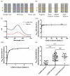
(A) SARS-CoV-2 Neutralizing Antibody Rapid Test Cassette (coupled with a spectrum-based reader) loaded with different COVID-19-confirmed cases. Control line region shown as an orange dotted line; test line region shown as a yellow dotted line. (B) SARS-CoV-2 Neutralizing Antibody Rapid Test Cassette (coupled with a spectrum-based reader) loaded with samples from different vaccinated individuals. Control line region shown as an orange dotted line; test line region shown as a yellow dotted line. (C) The reflectance spectra of a mild case, with the reflectance spectra of the control line region shown as a black solid line; the reflectance spectra of the test line region shown as a red solid line. The x-axis is the unit of wavelength (nm). The y-axis is the arbitrary unit (AU). (D1) Testing the NAb value based on the lateral flow immunoassay with a dilution series of a severe case patient’s serum. Curve fit by the Hill equation (R-squared = 0.9582), and (D2) is part of (D1) (R-squared = 0.9649). (E) Comparison of the NAb value based on the lateral flow immunoassay from healthy people (n = 10), COVID-19 patients (n = 30), and vaccinated people (n = 16). **** p < 0.0001; NS indicates no significant difference (p > 0.05).
Figure 3
Figure 3
(A) Comparison of the lateral flow immunoassay and ELISA for the neutralizing antibody using diluent series serum from a severe case (Rho = 0.9818, p-value < 0.0001). The x-axis is the unit of percent inhibition based on ELISA. The y-axis is the unit of NAb value based on the lateral flow immunoassay. (B) Comparison of the lateral flow immunoassay and ELISA for the neutralizing antibody using serum from patients, including 26 mild cases and 4 severe cases (n = 30) (Rho = 0.9288, p-value < 0.0001). The x-axis is the unit of percent inhibition based on ELISA. The y-axis is the unit of NAb value based on the lateral flow immunoassay. (C) Bland and Altman plot. The ratio between the neutralizing antibody based on the lateral flow immunoassay and ELISA in relation to the mean of the two measurements (n = 30). Green lines indicate the limits of agreement (±1.96 standard deviation). (D) ROC curve of the neutralizing antibody concentration in COVID-19 cases based on the lateral flow immunoassay and ELISA (n = 30) (AUC = 0.989, p < 0.001).
Figure 4
Figure 4
(A) Comparison of the lateral flow immunoassay and ELISA for the neutralizing antibody using plasma from vaccinated patients (Rho = 0.9818, p-value < 0.0001). The x-axis is the unit of percent inhibition based on ELISA. The y-axis is the unit of NAb value based on the lateral flow immunoassay. (B) Bland and Altman plot. The ratio between the neutralizing antibody based on the lateral flow immunoassay and ELISA in relation to the mean of the two measurements (n = 16). Green lines indicate the limits of agreement (±1.96 standard deviation). (C) ROC curve of the neutralizing antibody concentration in COVID-19-vaccinated cases based on the lateral flow immunoassay and ELISA (n = 16) (AUC = 0.933, p < 0.001).
Figure 5
Figure 5
(A) Correlation between the SARS-CoV-2 spike protein IgG antibody and neutralizing antibody from ELISA (Rho = 0.5724, p-value = 0.0035). The x-axis is the percent inhibition of the neutralizing antibody. The y-axis is the cut-off index for the SARS-CoV-2 spike protein IgG antibody. (B) Correlation between the human total IgG antibody and neutralizing antibody from ELISA. The x-axis is the unit of percent inhibition of the neutralizing antibody. The y-axis is the amount of total IgG antibody (ng/mL). (C) Neutralizing antibody tested from a saliva sample. We labeled the negative percent inhibition as zero. The x-axis is the saliva sample number. The y-axis is the unit of percent inhibition of the neutralizing antibody. The red dotted line represents the 30% inhibition based on ELISA, which is the cut-off value (positive or negative) of this assay.

References

    1. WHO WHO Statement Regarding Cluster of Pneumonia Cases in Wuhan, China. [(accessed on 7 January 2022)]. Available online: https://www.who.int/china/news/detail/09-01-2020-who-statement-regarding....
    1. WHO Coronavirus (COVID-19) Dashboard. [(accessed on 18 April 2021)]. Available online: https://covid19.who.int/
    1. Suthar M.S., Zimmerman M.G., Kauffman R.C., Mantus G., Linderman S.L., Hudson W.H., Vanderheiden A., Nyhoff L., Davis C.W., Adekunle O., et al. Rapid Generation of Neutralizing Antibody Responses in COVID-19 Patients. Cell Rep. Med. 2020;1:100040. doi: 10.1016/j.xcrm.2020.100040. - DOI - PMC - PubMed
    1. Wu F., Wang A., Liu M., Wang Q., Chen J., Xia S., Ling Y., Zhang Y., Xun J., Lu L., et al. Neutralizing antibody responses to SARS-CoV-2 in a COVID-19 recovered patient cohort and their implications. medRxiv. 2020 doi: 10.2139/ssrn.3566211. preprint . - DOI
    1. Wang X., Guo X., Xin Q., Pan Y., Hu Y., Li J., Chu Y., Feng Y., Wang Q. Neutralizing Antibody Responses to Severe Acute Respiratory Syndrome Coronavirus 2 in Coronavirus Disease 2019 Inpatients and Convalescent Patients. Clin. Infect. Dis. 2020;71:2688–2694. doi: 10.1093/cid/ciaa721. - DOI - PMC - PubMed