Case Reports
. 2022 Feb 16;10(2):302.
doi: 10.3390/vaccines10020302.

New-Onset Kidney Diseases after COVID-19 Vaccination: A Case Series

Affiliations
Case Reports

New-Onset Kidney Diseases after COVID-19 Vaccination: A Case Series

Jeong-Hoon Lim et al. Vaccines (Basel). .

Abstract

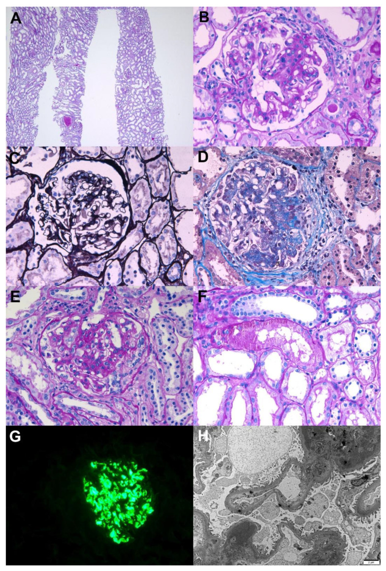
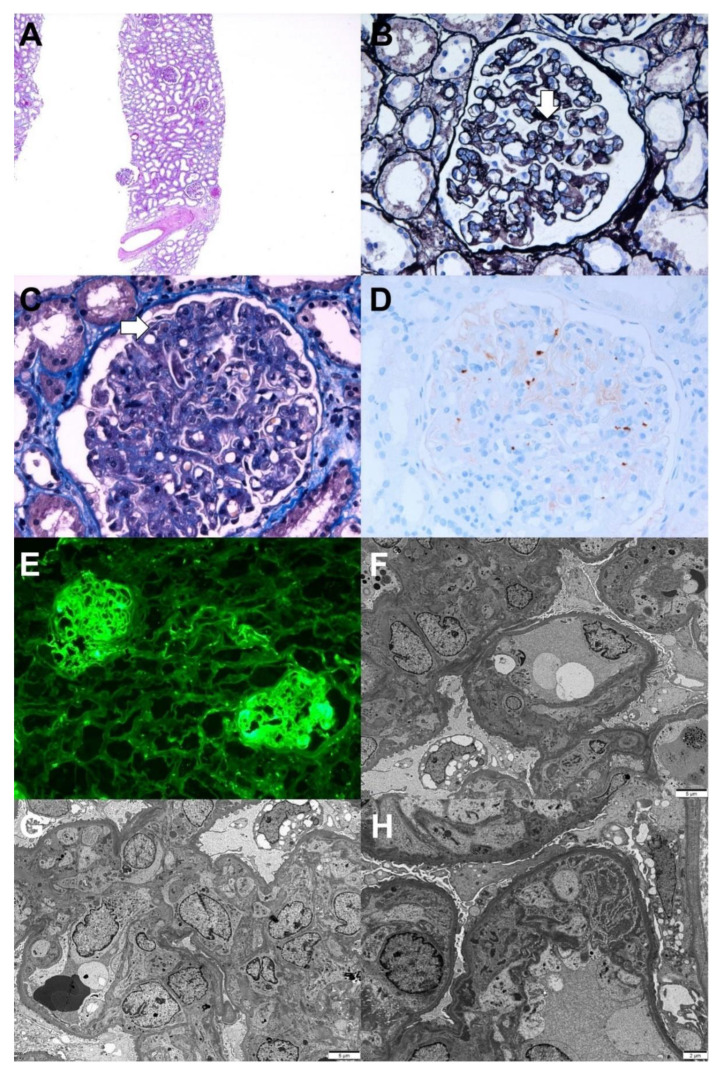
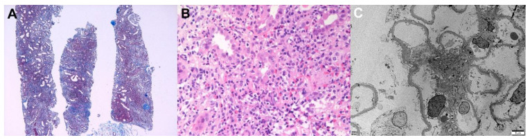
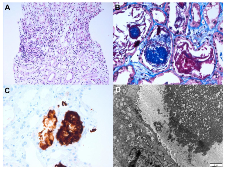
Various vaccines against COVID-19 have been developed and proven to be effective, but their side effects, especially on kidney function, are not yet known in detail. In this study, we report the clinical courses and histopathologic findings of new-onset kidney diseases after COVID-19 vaccination as confirmed via kidney biopsy. Five patients aged 42 to 77 years were included in this study, and baseline kidney function was normal in all patients. The biopsy-proven diagnosis indicated newly developed kidney diseases: (1) IgA nephropathy presenting with painless gross hematuria, (2) minimal change disease presenting with nephrotic syndrome, (3) thrombotic microangiopathy, and (4) two cases of acute tubulointerstitial nephritis presenting with acute kidney injury. Individualized treatment was applied as per disease severity and underlying pathology, and the treatment outcomes of all patients were improved. Since this is not a controlled study, the specific pathophysiologic link and causality between the incidence of kidney diseases and COVID-19 vaccination are difficult to confirm. However, clinicians need to consider the possibility that kidney diseases may be provoked by vaccines in patients who have renal symptoms.

Keywords: COVID-19; IgA nephropathy; kidney biopsy; kidney disease; minimal change disease; thrombotic microangiopathy; tubulointerstitial nephritis; vaccination.


Conflict of interest statement

The authors declare no conflict of interest.

Figures

Figure 1
Figure 1
Pathological findings of IgA nephropathy. Light microscopy: (A) In low magnification, no tubular atrophy or interstitial fibrosis was present (periodic acid–Schiff, ×40). (B) The glomerulus shows mesangial hypercellularity with increased mesangial matrix (periodic acid–Schiff, ×400). (C) The glomerulus shows endocapillary hypercellularity with focal loss of capillary penetrations (periodic acid methenamine silver, ×400). (D) The glomerulus shows segmental glomerulosclerosis (Masson’s trichrome, ×400). (E) The glomerulus shows cellular crescent with endocapillary hypercellularity (periodic acid–Schiff, ×400). (F) The picture shows protein reabsorption vacuoles in tubules, which is evidence of proteinuria (periodic acid–Schiff, ×400). Immunofluorescence: (G) Strong IgA mesangial positivity in the immunofluorescence stain (×400). Electron microscopy: (H) Numerous large electron-dense deposits in the mesangial areas with focal foot process effacement in the capillary surface were observed (×6000, 80 kv).
Figure 2
Figure 2
Pathologic findings of minimal change disease. Light microscopy: (A) In low magnification, no tubular atrophy or interstitial fibrosis was present (hematoxylin and eosin, ×40). (B) The picture shows a normally appearing glomerulus with protein reabsorption granules in tubules (hematoxylin and eosin, ×400). Electron microscopy: (C) Diffuse podocyte foot process effacement with microvillous transformation without electron-dense deposits were observed. The thickness of glomerular basememt membrane was normal (×2500, 80 kv).
Figure 3
Figure 3
Pathologic findings of chronic thrombotic microangiopathy. Light microscopy: (A) In low magnification, no tubular atrophy or interstitial fibrosis was present (hematoxylin and eosin, ×40). (B) The picture shows the duplication of the capillary basement membrane (arrow) (periodic acid methenamine silver, ×400). (C) The glomerular lumina were occluded due to the thickening of the glomerular capillary wall with a few fibrin thrombi (arrow) (Masson’s trichrome, ×400). (D) Immunohistochemical stain for CD61 (platelet glycoprotein GPIIIa) shows positivity against platelet thrombi (CD61, ×400). Immunofluorescence: (E) Strong positive staining for fibrinogen in the glomerulus was observed(×100). Electron microscopy: (F) Glomerular basement membrane duplication with cellular interpositions was observed (×3000, 80 kv). (G) Glomerular endothelial swelling and hypertrophy with occlusion of the lumens (glomerular endotheliosis) and diffuse foot process effacements along the capillary surfaces were observed (×3000, 80 kv). (H) Deposition of fibrinogen in the glomerular intracapillary area with entrapped cellular debris was noticed (×5000, 80 kv).
Figure 4
Figure 4
Pathologic findings of acute tubulointerstitial nephritis (patient 1). Light microscopy: (A) Massive interstitial inflammatory infiltrates with fibrosis (Masson’s trichrome, ×40) and (B) Mixed inflammatory cells infiltrated the tubular epithelium (tubulitis) and interstitium were observed. Large numbers of mononuclear cells, neutrophils, eosinophils, and plasma cells were present (hematoxylin and eosin, ×400). Electron microscopy: (C) The glomerulus showed a relatively normal appearance. No electron-dense deposits with a normal thickness of basement membrane and intact foot processes were present (×2500, 80 kv).
Figure 5
Figure 5
Pathologic findings of acute tubulointerstitial nephritis (patient 2). Light microscopy: (A) Mixed inflammatory cells infiltrated the tubular epithelium (tubulitis) and interstitium. Large numbers of mononuclear cells, neutrophils, eosinophils, and plasma cells were present (hematoxylin and eosin, ×400). (B) Acute tubular injury with red granular casts and string-like appearing casts was observed(Masson’s trichrome, ×400). (C) The casts show strong myoglobin positivity in the immunohistochemical stain (myoglobin, ×400). Electron microscopy: (D) The tubular casts have electron dense granules (×8000, 80 kv).

Similar articles

Cited by

References

    1. Pilishvili T., Gierke R., Fleming-Dutra K.E., Farrar J.L., Mohr N.M., Talan D.A., Krishnadasan A., Harland K.K., Smithline H.A., Hou P.C., et al. Effectiveness of mRNA Covid-19 vaccine among U.S. Health care personnel. N. Engl. J. Med. 2021;385:e90. doi: 10.1056/NEJMoa2106599. - DOI - PMC - PubMed
    1. Beatty A.L., Peyser N.D., Butcher X.E., Cocohoba J.M., Lin F., Olgin J.E., Pletcher M.J., Marcus G.M. Analysis of COVID-19 vaccine type and adverse effects following vaccination. JAMA Netw. Open. 2021;4:e2140364. doi: 10.1001/jamanetworkopen.2021.40364. - DOI - PMC - PubMed
    1. Lim J.H., Han M.H., Kim Y.J., Kim M.S., Jung H.Y., Choi J.Y., Cho J.H., Kim C.D., Kim Y.L., Park S.H. New-onset nephrotic syndrome after Janssen COVID-19 vaccination: A case report and literature review. J. Korean Med. Sci. 2021;36:e218. doi: 10.3346/jkms.2021.36.e218. - DOI - PMC - PubMed
    1. Klomjit N., Alexander M.P., Fervenza F.C., Zoghby Z., Garg A., Hogan M.C., Nasr S.H., Minshar M.A., Zand L. COVID-19 vaccination and glomerulonephritis. Kidney Int. Rep. 2021;6:2969–2978. doi: 10.1016/j.ekir.2021.09.008. - DOI - PMC - PubMed
    1. Bomback A.S., Kudose S., D’Agati V.D. De novo and relapsing glomerular diseases after COVID-19 vaccination: What do we know so far? Am. J. Kidney Dis. 2021;78:477–480. doi: 10.1053/j.ajkd.2021.06.004. - DOI - PMC - PubMed

Publication types

LinkOut - more resources