. 2022 Feb 15;11(4):527.
doi: 10.3390/plants11040527.

Jasmonate-Dependent Response of the Flower Abscission Zone Cells to Drought in Yellow Lupine

Affiliations

Jasmonate-Dependent Response of the Flower Abscission Zone Cells to Drought in Yellow Lupine

Agata Kućko et al. Plants (Basel). .

Abstract

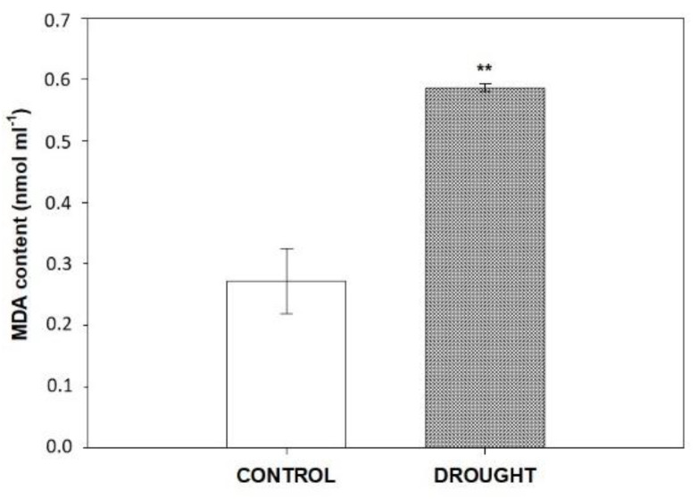
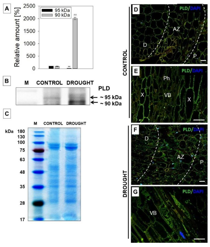
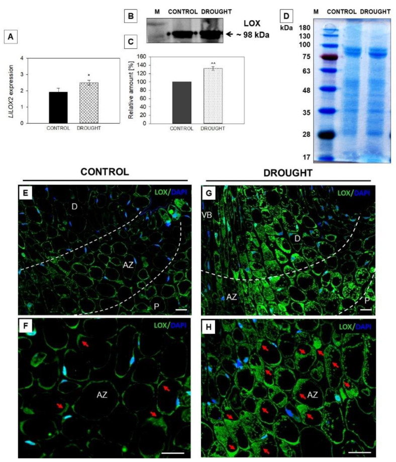
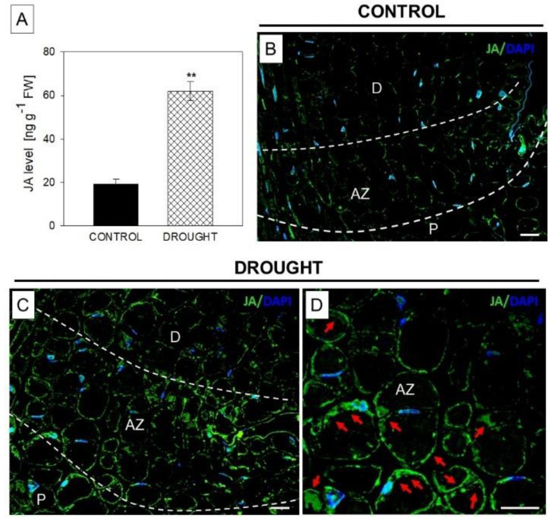
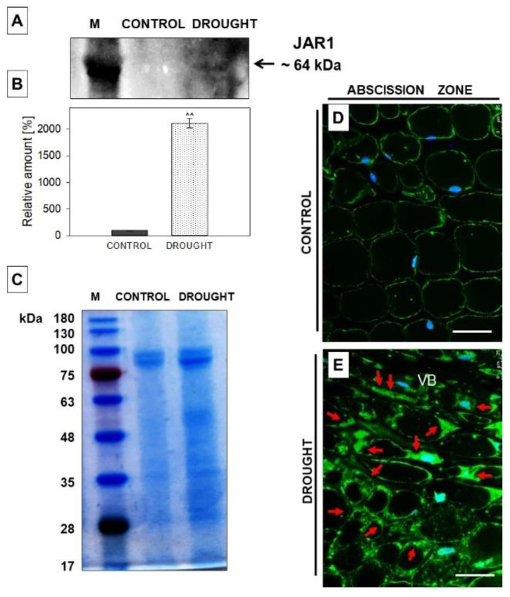
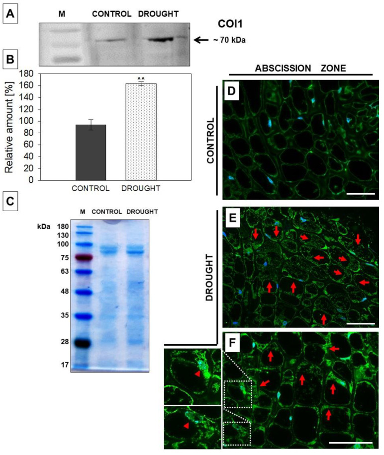
Lipid membranes, as primary places of the perception of environmental stimuli, are a source of various oxygenated polyunsaturated fatty acids-oxylipins-functioning as modulators of many signal transduction pathways, e.g., phytohormonal. Among exogenous factors acting on plant cells, special attention is given to drought, especially in highly sensitive crop species, such as yellow lupine. Here, we used this species to analyze the contribution of lipid-related enzymes and lipid-derived plant hormones in drought-evoked events taking place in a specialized group of cells-the flower abscission zone (AZ)-which is responsible for organ detachment from the plant body. We revealed that water deficits in the soil causes lipid peroxidation in these cells and the upregulation of phospholipase D, lipoxygenase, and, concomitantly, jasmonic acid (JA) strongly accumulates in AZ tissue. Furthermore, we followed key steps in JA conjugation and signaling under stressful conditions by monitoring the level and tissue localization of enzyme providing JA derivatives (JASMONATE RESISTANT1) and the JA receptor (CORONATINE INSENSITIVE1). Collectively, drought-triggered AZ activation during the process of flower abscission is closely associated with the lipid modifications, leading to the formation of JA, its conjugation, and induction of signaling pathways.

Keywords: CORONATINE INSENSITIVE1; JASMONATE RESISTANT1; abscission zone; drought; flower abscission; jasmonates; jasmonic acid; lipoxygenase; phospholipase D.


Conflict of interest statement

The authors declare no conflict of interest.

Figures

Figure 1
Figure 1
Effect of soil drought on the malondialdehyde (MDA) level in the flower abscission zone (AZ) of Lupinus luteus. Flower AZ was excised from plants subjected to 2 weeks of drought (25% water holding capacity, WHC) or lupines growing under optimal moisture (70% WHC, control). Results are presented as means ± SE (n = 3). Asterisks indicate a significant difference (Student’s t-test: ** p < 0.01).
Figure 2
Figure 2
The level of phospholipase D (PLD) in the flower abscission zone (AZ) of Lupinus luteus is strongly affected by soil drought. For the analyses, flowers’ AZ fragments were collected on the 48th day of cultivation from drought-treated lupines (25% water holding capacity, WHC) or control plants (70% WHC). For details, see the Material and Methods section. Western blot analysis was performed with an anti-PLD antibody and revealed the presence of two reactive bands of ~90 kDa and 95 kDa (B). These bands were scanned, and densitometry data were generated (100% was set for the control (A). Results on the chart are presented as means ± SE. A significant difference in AZ from drought-treated plants in comparison to control for the isoform of ~95 kDa are ** p < 0.01, while for the isoform of ~90 kDa are ^^ p < 0.01. Coomassie brilliant blue-stained SDS-PAGE gel (C). Molecular mass marker (M), sizes are shown in kDa. Immunolocalization of PLD in flower AZ of control (D,E) and drought-stressed lupines (F,G). Green fluorescence indicates the presence of PLD, whereas blue labeling corresponds to nuclei stained with DAPI. AZ region is marked by white dotted lines. Images (E,G) are magnified regions of the central area of AZ containing vascular tissues from control and stressed plants, respectively. Abbreviations: AZ—abscission zone; D—distal region of AZ; P—proximal region of AZ; Ph—phloem vessels; VB—vascular bundle, X—xylem vessels. Bars = 40 µM.
Figure 3
Figure 3
Lipoxygenase (LOX) is regulated by drought in the flower abscission zone (AZ) of Lupinus luteus. Analyses were carried out on flower AZs collected from 48 day-old lupines cultivated under drought (25% water holding capacity, WHC) or control (70% WHC) conditions. See Material and Methods for the details. Plant tissues were used for the expression analysis of LlLOX2 (A). Asterix indicates a significant difference with * p < 0.05. The isoform of LOX of ~98 kDa was obtained on the nitrocellulose membrane in the Western blot reaction with an anti-LOX antibody (B). A band was scanned and quantitative densitometric analysis was made (C, 100% was set for the control). ^^ p < 0.01 indicates a significant difference. SDS-PAGE electrophoresis gel stained with Coomassie Brilliant Blue (D). Sizes of molecular mass marker (M) are shown in kDa. Immunodetection of LOX in the flower’ AZ of control (E,F) and drought-stressed (G,H) plants. Green fluorescence indicates LOX accumulation (marked by red arrows), while blue labeling corresponds to nuclei stained with DAPI. AZ area is marked by white dotted lines. Images F and H are magnified regions of AZ from control and stressed tissues. Abbreviations: AZ—abscission zone; D—distal region of AZ; P—proximal region of AZ; VB—vascular bundle. Bars = 40 µM.
Figure 4
Figure 4
Drought upregulates the level of jasmonic acid (JA) in the abscission zone (AZ) of yellow lupine flowers. AZ fragments were collected for these analyses from 48 day-old plants subjected to drought (25% water holding capacity, WHC) or lupines cultivated in the soil of optimal moisture (70% WHC, control). Details in the Material and Methods section. The level of JA was examined using GC-MS (A). A significant difference is ** p < 0.01. Immunolocalization of JA in the AZ of control (B) or stressed (C,D) plants. Green fluorescence is related to JA localization (indicated by red arrows), whereas blue color corresponds to DAPI-stained nuclei. AZ area is marked by white dotted lines (B,C). (D) image is magnified AZ area presented on C. Abbreviations: AZ—abscission zone; D—distal region of AZ; P—proximal region of AZ. Bars = 40 µM.
Figure 5
Figure 5
JASMONATE RESISTANT1 (JAR1) is induced in the abscission zone (AZ) cells of yellow lupine flowers in response to soil drought. AZ from flowers collected from 48 day-old plants growing under optimal soil moisture (70% water holding capacity, WHC) was a control, while the “drought” variant was AZ excised from 48 day-old plants subjected to drought (25% WHC), as written precisely in the Material and Methods section. Western blot analysis was performed with a JAR1 antibody and revealed the presence of a reactive band of ~64 kDa (A). The membrane was scanned, densitometry analysis was performed (100% was set for the control), and presented in (B). Protein samples separated by SDS-PAGE and stained with Coomassie Brilliant Blue (C). Protein marker (M) is displayed on the left and sizes in kDa are provided. Immunolocalization of JAR1 in flower AZ cells during soil water deficit (E) and under control conditions (D). Green fluorescence indicates JAR1 localization (marked by red arrows). Blue labeling corresponds to DAPI-stained nuclei. AZ region is restricted by white dotted lines. Abbreviation: VB—vascular bundle. Bars = 40 µM. A significant difference is ^^ p < 0.01.
Figure 6
Figure 6
The content of coronatine insensitive 1 (COI1) in the flower abscission zone (AZ) of Lupinus luteus strongly increased under soil water deficit. Flower AZ was harvested from plants growing in the soil of optimal moisture (70% water holding capacity, WHC, control) or drought-stressed lupines (25% WHC). Detailed description in the Material and Methods section. Proteins were isolated and subjected to Western blot analysis with an anti-COI1 antibody, which revealed the presence of a band of ~70 kDa (A). Quantitative densitometry analysis was made and presented in (B) (100% was set for the control). ^^ p < 0.01 indicates a significant difference. Protein samples were separated by SDS-PAGE and stained with Coomassie Brilliant Blue (C). Mass marker (M) is provided on the left, sizes of proteins are displayed in kDa. COI1 antibody was used for immunolocalization analysis performed on control (D) and drought-stressed (E,F) tissues of AZ from flowers. A green fluorescence signal, indicated by red arrows, corresponds to the presence of COI1. Nuclei were detected by DAPI (blue color). Enlarged cells of stressed AZ are provided on the left side of the (F) image. These are magnified regions marked by white boxes and show colocalization of CO1 with nuclei (red arrowheads). Bars = 40 µM.

Similar articles

Cited by

References

    1. Huang H., Liu B., Liu L., Song S. Jasmonate action in plant growth and development. J. Exp. Bot. 2017;68:1349–1359. doi: 10.1093/jxb/erw495. - DOI - PubMed
    1. Ku Y.-S., Sintaha M., Cheung M.-Y., Lam H.-M. Plant hormone signaling crosstalks between biotic and abiotic stress responses. Int. J. Mol. Sci. 2018;19:3206. doi: 10.3390/ijms19103206. - DOI - PMC - PubMed
    1. Li M., Yu G., Cao C., Liu P. Metabolism, signaling, and transport of jasmonates. Plant Commun. 2021;2:100231. doi: 10.1016/j.xplc.2021.100231. - DOI - PMC - PubMed
    1. He Y., Fukushige H., Hildebrand D.F., Gan S. Evidence supporting a role of jasmonic acid in Arabidopsis leaf senescence. Plant Physiol. 2002;128:876–884. doi: 10.1104/pp.010843. - DOI - PMC - PubMed
    1. Kim J., Chang C., Tucker M.L. To grow old: Regulatory role of ethylene and jasmonic acid in senescence. Front. Plant Sci. 2015;6:20. doi: 10.3389/fpls.2015.00020. - DOI - PMC - PubMed

LinkOut - more resources