. 2022 Feb 14;12(4):641.
doi: 10.3390/nano12040641.

Antibacterial and Fluorescence Staining Properties of an Innovative GTR Membrane Containing 45S5BGs and AIE Molecules In Vitro

Affiliations

Antibacterial and Fluorescence Staining Properties of an Innovative GTR Membrane Containing 45S5BGs and AIE Molecules In Vitro

Yu-Wen Wei et al. Nanomaterials (Basel). .

Abstract

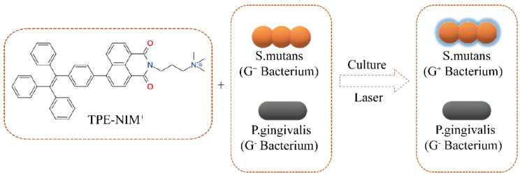
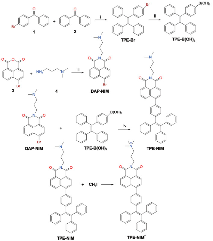
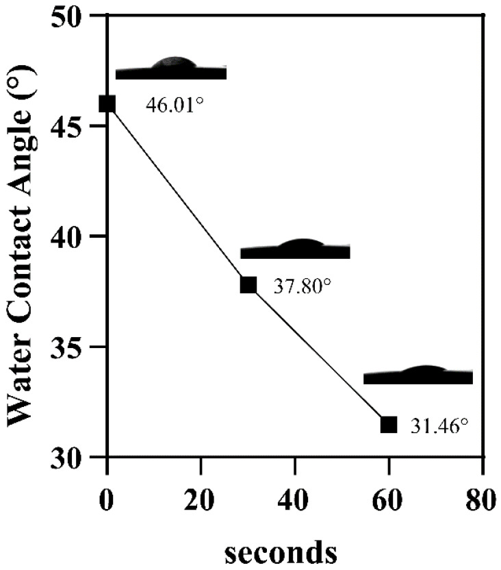
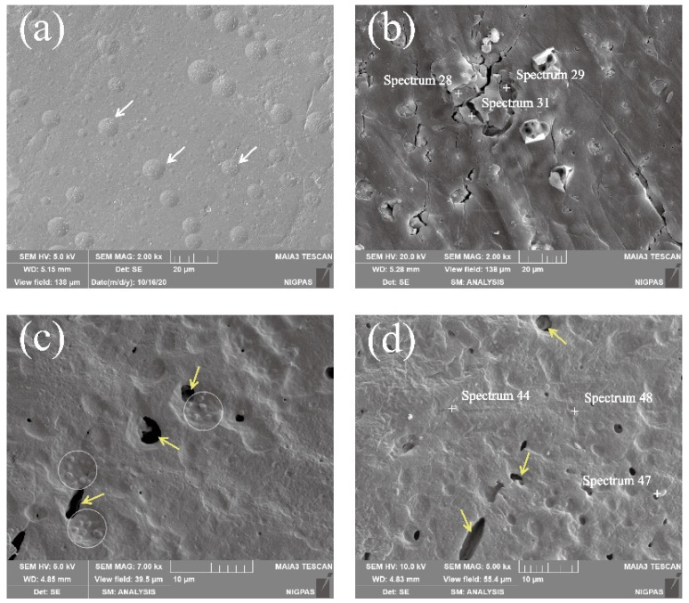
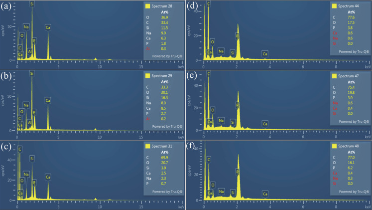
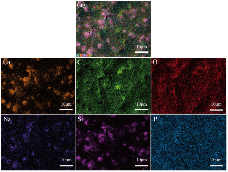
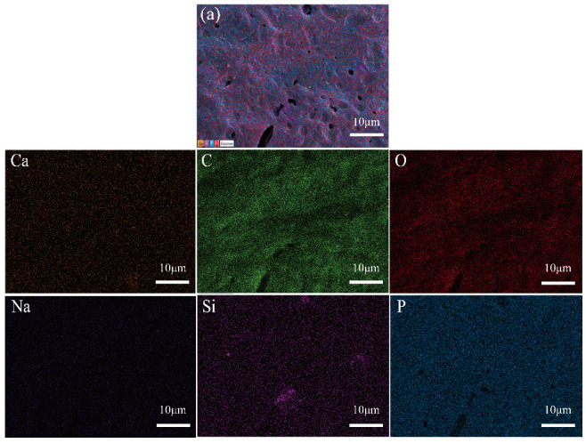
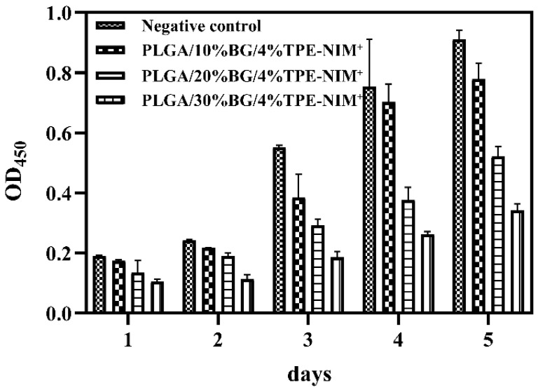
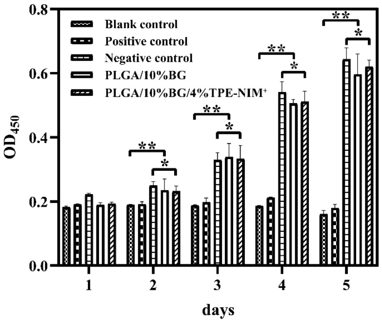
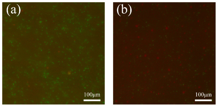
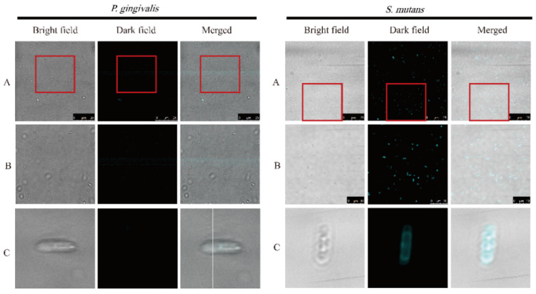
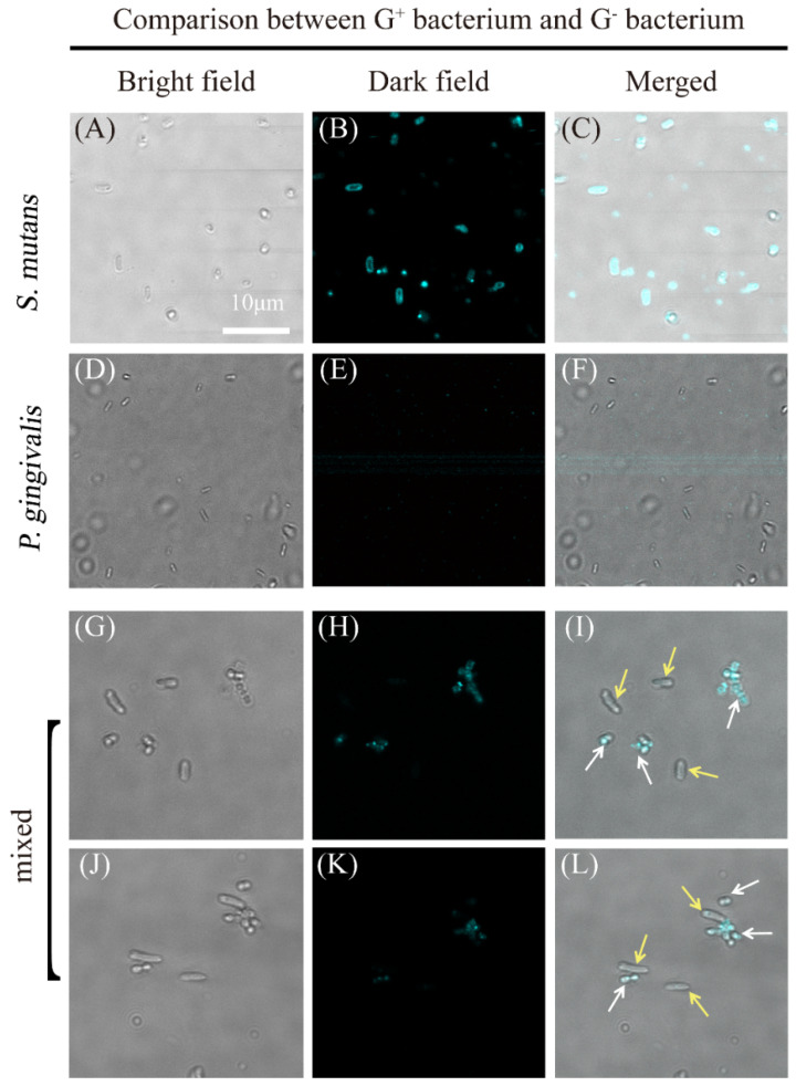
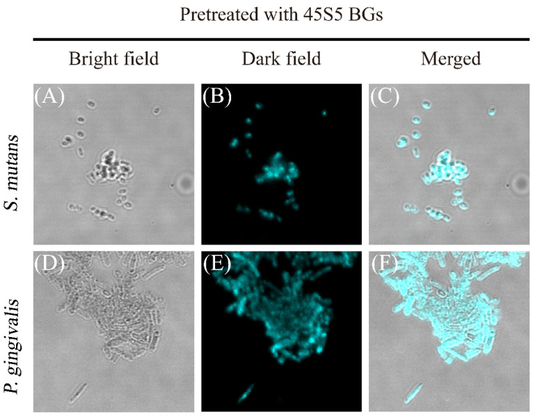
This study aimed to add two functional components-antibacterial 45S5BGs particles and AIE nanoparticles (TPE-NIM+) with bioprobe characteristics-to the guided tissue regeneration (GTR) membrane, to optimize the performance. The PLGA/BG/TPE-NIM+ membrane was synthesized. The static water contact angle, morphologies, and surface element analysis of the membrane were then characterized. In vitro biocompatibility was tested with MC3T3-E1 cells using CCK-8 assay, and antibacterial property was evaluated with Streptococcus mutans and Porphyromonas gingivalis by the LIVE/DEAD bacterial staining and dilution plating procedure. The fluorescence staining of bacteria was observed by Laser Scanning Confocal Microscope. The results showed that the average water contact angle was 46°. In the cytotoxicity test, except for the positive control group, there was no significant difference among the groups (p > 0.05). The antibacterial effect in the PLGA/BG/TPE-NIM+ group was significantly (p < 0.01), while the sterilization rate was 99.99%, better than that in the PLGA/BG group (98.62%) (p < 0.01). Confocal images showed that the membrane efficiently distinguished G+ bacteria from G- bacteria. This study demonstrated that the PLGA/BG/TPE-NIM+ membrane showed good biocompatibility, efficient sterilization performance, and surface mineralization ability and could be used to detect pathogens in a simple, fast, and wash-free protocol.

Keywords: aggregation-induced emission nanoparticles; antibacterial; antibacterial differentiation; bioactive glass; bioprobe; guided tissue regeneration (GTR).


Conflict of interest statement

The authors declare no conflict of interest. The funders had no role in the design of the study; in the collection, analyses, or interpretation of data; in the writing of the manuscript; or in the decision to publish the results.

Figures

Scheme 1
Scheme 1
Molecular structures of TPE-NIM+ and a general representation of their adsorption onto the surfaces of G+ bacterium. Note that the TPE-NIM+ and the bacteria are not drawn in real scales.
Scheme 2
Scheme 2
Synthetic routes of TPE-NIM+. Reagents and conditions: (i) Zn, TiCl4, tetrahydrofuran (THF), 0–25 °C, 12 h, (ii) n-Butyllithium solution, trimethyl borate, THF, –78 °C, 3 h, (iii) C2H5OH, reflux, 2 h, and (iv) Pd(PPh3)4, Na2CO3, mixed solvent (toluene: ethanol: water), 90 °C, 12 h.
Figure 1
Figure 1
The Proton NMR of TPE-NIM+.
Figure 2
Figure 2
Images of the surface of the PLGA/BG/TPE-NIM+ membrane after putting a drop of distilled water on it. Measured water contact angle values and captured images at 0 s, 30 s, and 60 s.
Figure 3
Figure 3
SEM images of composite membranes before and after immersion in SBF. (a,b) SEM images of PLGA/10%BG/4%TPE-NIM+ membrane before immersion in SBF. White arrows show the exposed 45S5BGs particles. (b) Three fixed points (Spectrum 28.29.31) were selected randomly for EDS analysis. (c,d) SEM images of PLGA/10%BG/4%TPE-NIM+ membrane after immersion in SBF for three days: (c) White circles showed the embedded 45S5BGs particles. Yellow arrows showed the hollow structure; and (d) Yellow arrows show the hollow structure. Three fixed points (Spectrum 44.47.48) were selected randomly for EDS analysis.
Figure 4
Figure 4
EDS analysis of composite membranes before and after immersion. (ac) EDS images of PLGA/10%BG/4%TPE-NIM+ membrane without immersion in SBF. (df) EDS images of PLGA/10%BG/4%TPE-NIM+ membrane after three days of immersion in SBF.
Figure 5
Figure 5
EDS Elemental Analysis with ColorSEM Technology of the PLGA/10%BG/4%TPE-NIM+ membrane without immersion in SBF. (a) EDS image with all elements superimposed.
Figure 6
Figure 6
EDS Elemental Analysis with ColorSEM Technology of the PLGA/10%BG/4%TPE-NIM+ membrane after three days of immersion in SBF. (a) EDS image with all elements superimposed.
Figure 7
Figure 7
Cytotoxicity of control medium, PLGA/BG/TPE-NIM+ composite membranes, respectively, containing 10%, 20%, or 30% BG toward MC3T3-E1.
Figure 8
Figure 8
Cytotoxicity of control medium, PLGA/BG membrane and PLGA/BG/TPE-NIM+ membrane toward MC3T3-E1. (* p > 0.05 ** p < 0.05 vs. blank control group).
Figure 9
Figure 9
Live and Dead bacterial staining. (a) S. m bacteria liquid without the treatment with PLGA/BG/TPE-NIM+ composite membrane. (b) S. m bacteria liquid with the treatment with PLGA/BG/TPE-NIM+ composite membrane for 24 h.
Figure 10
Figure 10
Confocal images of P. gingivalis (left: AC) and S. mutans (right: AC) after treatment with PLGA/10%BG/4%TPE-NIM+ membrane extract (without 45S5BGs) for 15 min.
Figure 11
Figure 11
Confocal images of S. mutans (AC) and P. gingivalis (DF) after treatment with PLGA/10%BG/4%TPE-NIM+ membrane extract (without 45S5BGs) for 15 min. (GL) Selective staining of S. mutans by TPE-NIM+. White arrow indicates stained S. mutans and yellow arrows indicate non-stained P. gingivalis.
Figure 12
Figure 12
Confocal images of S. mutans (AC) and P. gingivalis (DF) after treatment with 45S5BGs and PLGA/10%BG/4%TPE-NIM+ membrane extract (with 45S5BGs), each for 15 min.

Similar articles

Cited by

References

    1. Kinane D.F., Stathopoulou P.G., Papapanou P.N. Periodontal diseases. Nat. Rev. Dis. Primers. 2017;3:17038. doi: 10.1038/nrdp.2017.38. - DOI - PubMed
    1. Schwarz F., Derks J., Monje A., Wang H.L. Peri-implantitis. J. Periodontol. 2018;89((Suppl. 1)):S267–S290. doi: 10.1002/JPER.16-0350. - DOI - PubMed
    1. Machtei E.E., Cho M.I., Dunford R., Norderyd J., Zambon J.J., Genco R.J. Clinical, microbiological, and histological factors which influence the success of regenerative periodontal therapy. J. Periodontol. 1994;65:154–161. doi: 10.1902/jop.1994.65.2.154. - DOI - PubMed
    1. Carcuac O., Derks J., Charalampakis G., Abrahamsson I., Wennström J., Berglundh T. Adjunctive Systemic and Local Antimicrobial Therapy in the Surgical Treatment of Peri-implantitis: A Randomized Controlled Clinical Trial. J. Dent. Res. 2016;95:50–57. doi: 10.1177/0022034515601961. - DOI - PubMed
    1. Renvert S., Roos-Jansåker A.M., Claffey N. Non-surgical treatment of peri-implant mucositis and peri-implantitis: A literature review. J. Clin. Periodontol. 2008;35:305–315. doi: 10.1111/j.1600-051X.2008.01276.x. - DOI - PubMed

LinkOut - more resources