. 2022 Jan 27;11(2):173.
doi: 10.3390/pathogens11020173.

The Coxsackievirus and Adenovirus Receptor Has a Short Half-Life in Epithelial Cells

Affiliations

The Coxsackievirus and Adenovirus Receptor Has a Short Half-Life in Epithelial Cells

Poornima Kotha Lakshmi Narayan et al. Pathogens. .

Abstract

The coxsackievirus and adenovirus receptor (CAR) is an essential cellular protein that is involved in cell adhesion, cell signaling, and viral infection. The 8-exon encoded isoform (CAREx8) resides at the apical surface of polarized epithelia, where it is accessible as a receptor for adenovirus entering the airway lumen. Given its pivotal role in viral infection, it is a target for antiviral strategies. To understand the regulation of CAREx8 and determine the feasibility of receptor downregulation, the half-life of total and apical localized CAREx8 was determined and correlated with adenovirus transduction. Total and apical CAREx8 has a relatively short half-life of approximately 2 h. The half-life of apical CAREx8 correlates well with adenovirus transduction. These results suggest that antiviral strategies that aim to degrade the primary receptor for apical adenovirus infection will be effective within a relatively short time frame after application.

Keywords: coxsackievirus and adenovirus receptor; half-life; human adenovirus; polarized epithelia.


Conflict of interest statement

The authors declare no conflict of interest. The funders had no role in the design of the study; in the collection, analyses, or interpretation of data; in the writing of the manuscript, or in the decision to publish the results.

Figures

Figure 1
Figure 1
CAREx8 has a half-life of approximately 2 h. MDCK cells that stably expressed FLAG-tagged CAREx8 under the control of a doxycycline-sensitive promoter were seeded in a plastic 12-well dish in FBS tet(−) media. Cells were induced with 100 ng/mL doxycycline for 24 h. The doxycycline-containing media were then removed and replaced with standard FBS tet(−) culture media. (A). Cells were lysed at indicated time points before and after removal of Dox and the lysates were analyzed by Western blot (WB) using anti-FLAG or actin-specific primary antibodies. (B). Cells were lysed at indicated time points post Dox removal and lysates were analyzed by WB. (C). CAREx8 bands were normalized to their corresponding actin bands and quantified using ImageJ. (D). MDCK-FLAG-CAREx8 were polarized on semipermeable membranes. At 24 h post Dox treatment, standard FBS tet(−) culture media were added and surface biotinylation was performed at indicated time points post Dox removal. Cells were then lysed and the lysates (actin) or isolated-biotinylated proteins (CAREx8, FLAG Ab) were analyzed via WB. (E). CAREx8 bands were normalized to their corresponding actin bands and quantified using ImageJ. Error bars represent SEM. (* p < 0.05; ** p < 0.01; One-Way ANOVA; N at least 3).
Figure 2
Figure 2
CAREx8 has a half-life of approximately 2 h. (A) Calu-3 cells were treated with 30 μg/mL cycloheximide. The cells were lysed at the times indicated post cycloheximide addition and analyzed by WB using a CAREx8-specific primary antibody (5678p) or actin (loading control). (B) Quantification of CAREx8 bands normalized to actin. Error bars represent SEM. (* p < 0.05; ** p < 0.01; *** p < 0.001; One-Way ANOVA; N at least 3).
Figure 3
Figure 3
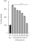
Adenovirus transduction decreases as CAREx8 degrades. MDCK-FLAG-CAREx8 were seeded and polarized in FBS tet(−) media. The cells were then treated with 100 ng/mL Dox for 24 h. At 24 h post treatment, the doxycycline-containing media were removed and replaced with standard FBS tet(−) culture media. At 4, 6, 8, 10, 12, and 24 h post dox removal, cells were transduced with AdV-β-Gal. AdV transduction was analyzed by β-galactosidase assay after 24 h. Error bars indicate standard error of the mean. (**** p < 0.0001; One-Way ANOVA; N = 3).

References

    1. Tregoning J.S., Schwarze J. Respiratory viral infections in infants: Causes, clinical symptoms, virology, and immunology. Clin. Microbiol. Rev. 2010;23:74–98. doi: 10.1128/CMR.00032-09. - DOI - PMC - PubMed
    1. Vareille M., Kieninger E., Edwards M.R., Regamey N. The airway epithelium: Soldier in the fight against respiratory viruses. Clin. Microbiol. Rev. 2011;24:210–229. doi: 10.1128/CMR.00014-10. - DOI - PMC - PubMed
    1. William S.M., Wold M.S.H. Adenoviruses. In: David M., Knipe P.M.H., editors. Fields Virology. Volume 2. Lippincott Williams and Wilkins; Philadelphia, PA, USA: 2007. pp. 2395–2436.
    1. Robinson C.M., Singh G., Lee J.Y., Dehghan S., Rajaiya J., Liu E.B., Yousuf M.A., Betensky R.A., Jones M.S., Dyer D.W., et al. Molecular evolution of human adenoviruses. Sci. Rep. 2013;3:1812. doi: 10.1038/srep01812. - DOI - PMC - PubMed
    1. Arnberg N. Adenovirus receptors: Implications for targeting of viral vectors. Trends Pharmacol. Sci. 2012;33:442–448. doi: 10.1016/j.tips.2012.04.005. - DOI - PubMed

LinkOut - more resources