. 2022 Feb 12;11(2):239.
doi: 10.3390/pathogens11020239.

Evaluation of Serological Markers in Alveolar Echinococcosis Emphasizing the Correlation of PET-CTI Tracer Uptake with RecEm18 and Echinococcus-Specific IgG

Affiliations

Evaluation of Serological Markers in Alveolar Echinococcosis Emphasizing the Correlation of PET-CTI Tracer Uptake with RecEm18 and Echinococcus-Specific IgG

Julian Frederic Hotz et al. Pathogens. .

Abstract

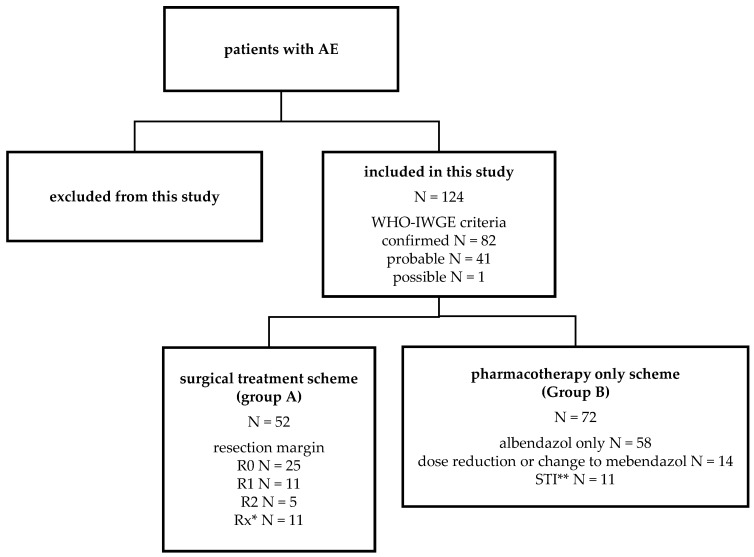
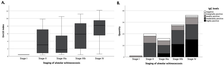
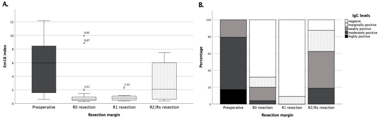
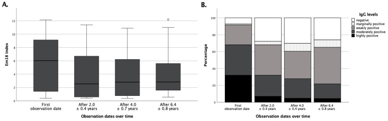
Human alveolar echinococcosis (AE), which is caused by the cestode Echinococcus (E.) multilocularis, is an epidemiologically relevant issue in modern medicine and still poses a diagnostic and therapeutic challenge. Since diagnosis mainly relies on imaging procedures and serological testing, we retrospectively and comparatively analyzed the performance of an Echinococcus IgG screening ELISA, whole serum IgE, and two specific confirmatory ELISA platforms using the defined E. multilocularis antigens Em2-Em18 (Em2+) and recombinant Em18 (recEm18). With special emphasis on the clinical usefulness of recEm18, we correlated the laboratory results with clinical characteristics and imaging findings in a large and well-characterized cohort of N = 124 AE patients, who were followed over several years after either surgical plus subsequent pharmacological treatment or pharmacotherapy alone. All patients had routinely received PET-CTI every two years. Our data reveal strong correlations for both Echinococcus IgG and recEm18 with tracer uptake in PET-CTI and parasitic lesion size and number, suggesting additional clinical usefulness of recEm18 for certain constellations only, while IgG and Em2+ still appear reasonable and sensitive screening methods for initial diagnosis of AE. With this study, we aim to contribute to further optimizing medical care of AE patients. For instance, it might be reasonable to consider the replacement of some PET-CTI follow-ups by imaging procedures with less radiation exposure or serological means alone. Further studies that clarify the correlation of serological markers with ultrasound criteria might be particularly useful, and further retrospective as well as prospective investigations are justified in this context.

Keywords: Echinococcus IgG; PET-CTI; alveolar echinococcosis; recEm18.


Conflict of interest statement

The authors declare no conflict of interest.

Figures

Figure 1
Figure 1
Grouping of patient collectives according to treatment schemes. Alveolar echinococcosis (AE) was classified as possible, probable, or confirmed according to the WHO–IWGE criteria [8]. * Unknown resection margin, ** structured treatment interruption.
Figure 2
Figure 2
Correlations of recEm18 indices and total Echinococcus IgG depending on the initial staging. Patients of group C (N = 101) were included. (Panel A) shows the stage-dependent recEm18 indices. Accordingly, (panel B) exhibits the number of semi-quantitative detections of Echinococcus IgG depending on the patients’ staging.
Figure 3
Figure 3
Levels of pre- and postoperative recEm18 indices depend on the resection margin. (Panel A) shows the recEm18 indices of group A both preoperatively and after resection depending on the resection margin, while (panel B) exhibits the percentage of semi-quantitative total Echinococcus IgG levels pre- and postoperatively according to the resection margin after surgical treatment. Serological controls were performed on average 1.9 ± 0.6 years after R0, 2.2 ± 0.2 years after R1, and 1.6 ± 2.3 years after R2/Rx resection.
Figure 4
Figure 4
RecEm18 index and Echinococcus IgG under benzimidazole therapy over time. (Panel A) shows boxplots for patients of group A at different instances of time during follow-up. The mean recEm18 indices at the respective time points were 5.38 ± 3.70 (median 6.03, N = 72) on the first appointment; 3.72 ± 3.40 (median 2.55, N = 72) after 2.0 ± 0.4 years; 3.72 ± 3.31 (median 2.80, N = 43) after 4.0 ± 0.7 years; and 4.16 ± 3.52 (median 2.83, N = 23) after 6.4 ± 0.8 years. (Panel B) shows the portions of semi-quantitative IgG levels over time for patients of group A. In total, 31.9% were highly positive, 6.94% negative (N = 72) on the first appointment; 6.9% highly positive, 27.8% negative (N = 72) after 2.0 ± 0.4 years; 4.65% highly positive, 30.2% negative (N = 43) after 4.0 ± 0.7 years; 4.4% highly positive, 26.1% negative (N = 23) after 6.4 ± 0.8 years, respectively.
Figure 5
Figure 5
Percentage of positive results of all parameters depending on different resection margins. The graph shows the percentage of positive test results of group A pre- and postoperatively according to the resection margin (R0, R1, and R2/Rx resection). Preoperatively, recEm18 index, IgG, and total IgE were measured in N = 29, while Em2+ ELISA results were collected for N = 27 patients. Postoperatively, all parameters were determined in parallel from all serum samples, so no differences in the respective number of test results came about.

References

    1. Woolsey I.D., Miller A.L. Echinococcus granulosus sensu lato and Echinococcus multilocularis: A review. Res. Vet. Sci. 2021;135:517–522. doi: 10.1016/j.rvsc.2020.11.010. - DOI - PubMed
    1. McManus D.P., Zhang W., Li J., Bartley P.B. Echinococcosis. Lancet. 2003;362:1295–1304. doi: 10.1016/S0140-6736(03)14573-4. - DOI - PubMed
    1. Gruener B., Kern P., Mayer B., Graeter T., Hillenbrand A., Barth T.E.F., Muche R., Henne-Bruns D., Kratzer W., Kern P. Comprehensive diagnosis and treatment of alveolar echinococcosis: A single-center, long-term observational study of 312 patients in Germany. GMS Infect. Dis. 2017;5:Doc01. doi: 10.3205/id000027. - DOI - PMC - PubMed
    1. Kern P., Wen H., Sato N., Vuitton D.A., Gruener B., Shao Y., Delabrousse E., Kratzer W., Bresson-Hadni S. WHO classification of alveolar echinococcosis: Principles and application. Parasitol. Int. 2006;55:S283–S287. doi: 10.1016/j.parint.2005.11.041. - DOI - PubMed
    1. Reinehr M., Micheloud C., Grimm F., Kronenberg P.A., Grimm J., Beck A., Nell J., Meyer zu Schwabedissen C., Furrer E., Muellhaupt B., et al. Pathology of echinococcosis: A morphologic and immunohistochemical study on 138 specimens with focus on the differential diagnosis between cystic and alveolar Echinococcosis. Am. J. Surg. Pathol. 2020;44:43–54. doi: 10.1097/PAS.0000000000001374. - DOI - PubMed

LinkOut - more resources