

The p97 Inhibitor UPCDC-30245 Blocks Endo-Lysosomal Degradation
- PMID: 35215314
- PMCID: PMC8880557
- DOI: 10.3390/ph15020204
The p97 Inhibitor UPCDC-30245 Blocks Endo-Lysosomal Degradation
Abstract
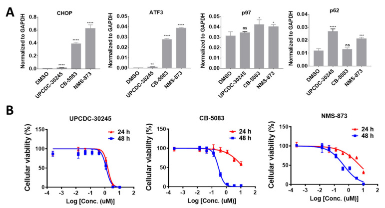
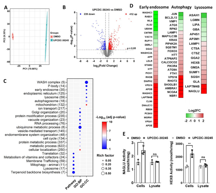
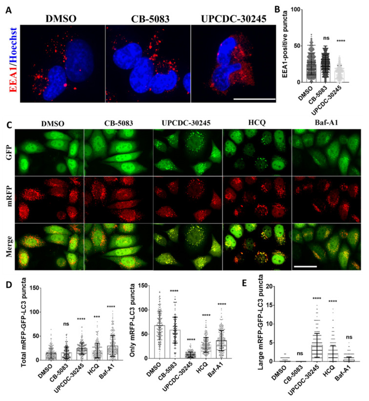
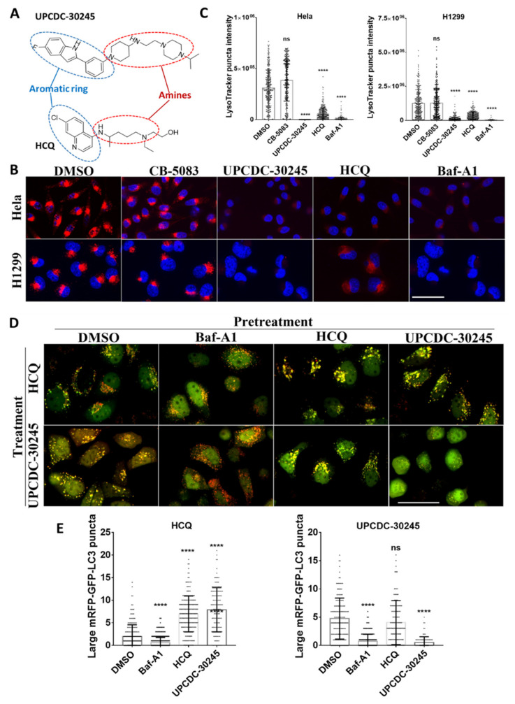
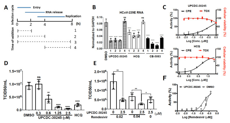
The diverse modes of action of small molecule inhibitors provide versatile tools to investigate basic biology and develop therapeutics. However, it remains a challenging task to evaluate their exact mechanisms of action. We identified two classes of inhibitors for the p97 ATPase: ATP competitive and allosteric. We showed that the allosteric p97 inhibitor, UPCDC-30245, does not affect two well-known cellular functions of p97, endoplasmic-reticulum-associated protein degradation and the unfolded protein response pathway; instead, it strongly increases the lipidated form of microtubule-associated proteins 1A/1B light chain 3B (LC3-II), suggesting an alteration of autophagic pathways. To evaluate the molecular mechanism, we performed proteomic analysis of UPCDC-30245 treated cells. Our results revealed that UPCDC-30245 blocks endo-lysosomal degradation by inhibiting the formation of early endosome and reducing the acidity of the lysosome, an effect not observed with the potent p97 inhibitor CB-5083. This unique effect allows us to demonstrate UPCDC-30245 exhibits antiviral effects against coronavirus by blocking viral entry.
Keywords: coronavirus; endo-lysosomal degradation; lysomotropic agents; p97 inhibitor; proteomics.
Conflict of interest statement
The authors declare no conflict of interest.
Figures
Similar articles
-
Allosteric p97 Inhibitors Can Overcome Resistance to ATP-Competitive p97 Inhibitors for Potential Anticancer Therapy.ChemMedChem. 2020 Apr 20;15(8):685-694. doi: 10.1002/cmdc.201900722. Epub 2020 Mar 23. ChemMedChem. 2020. PMID: 32162487 Free PMC article.
-
Development of p97 AAA ATPase inhibitors.Autophagy. 2011 Sep;7(9):1091-2. doi: 10.4161/auto.7.9.16489. Epub 2011 Sep 1. Autophagy. 2011. PMID: 21606684 Free PMC article.
-
Reversible inhibitor of p97, DBeQ, impairs both ubiquitin-dependent and autophagic protein clearance pathways.Proc Natl Acad Sci U S A. 2011 Mar 22;108(12):4834-9. doi: 10.1073/pnas.1015312108. Epub 2011 Mar 7. Proc Natl Acad Sci U S A. 2011. PMID: 21383145 Free PMC article.
-
VCP/p97/Cdc48, A Linking of Protein Homeostasis and Cancer Therapy.Curr Mol Med. 2017;17(9):608-618. doi: 10.2174/1566524018666180308111238. Curr Mol Med. 2017. PMID: 29521227 Review.
-
Toward an understanding of the Cdc48/p97 ATPase.F1000Res. 2017 Aug 3;6:1318. doi: 10.12688/f1000research.11683.1. eCollection 2017. F1000Res. 2017. PMID: 28815021 Free PMC article. Review.
Cited by
-
The functional importance of VCP to maintaining cellular protein homeostasis.Biochem Soc Trans. 2022 Oct 31;50(5):1457-1469. doi: 10.1042/BST20220648. Biochem Soc Trans. 2022. PMID: 36196920 Free PMC article. Review.
-
p97 Inhibitors Possessing Antiviral Activity Against SARS-CoV-2 and Low Cytotoxicity.Pharmaceuticals (Basel). 2025 Jan 19;18(1):131. doi: 10.3390/ph18010131. Pharmaceuticals (Basel). 2025. PMID: 39861192 Free PMC article.
-
A genome-scale drug discovery pipeline uncovers new therapeutic targets and a unique p97 allosteric binding site in Schistosoma mansoni.bioRxiv [Preprint]. 2025 Mar 15:2025.03.14.643303. doi: 10.1101/2025.03.14.643303. bioRxiv. 2025. PMID: 40161785 Free PMC article. Preprint.
-
Mechanism of allosteric inhibition of human p97/VCP ATPase and its disease mutant by triazole inhibitors.Commun Chem. 2024 Aug 9;7(1):177. doi: 10.1038/s42004-024-01267-3. Commun Chem. 2024. PMID: 39122922 Free PMC article.
References
-
- Anderson D.J., Le Moigne R., Djakovic S., Kumar B., Rice J., Wong S., Wang J., Yao B., Valle E., Kiss von Soly S., et al. Targeting the AAA ATPase p97 as an Approach to Treat Cancer through Disruption of Protein Homeostasis. Cancer Cell. 2015;28:653–665. doi: 10.1016/j.ccell.2015.10.002. - DOI - PMC - PubMed
-
- Her N.G., Toth J.I., Ma C.T., Wei Y., Motamedchaboki K., Sergienko E., Petroski M.D. p97 Composition Changes Caused by Allosteric Inhibition Are Suppressed by an On-Target Mechanism that Increases the Enzyme’s ATPase Activity. Cell. Chem. Biol. 2016;23:517–528. doi: 10.1016/j.chembiol.2016.03.012. - DOI - PMC - PubMed
-
- Burnett J.C., Lim C., Peyser B.D., Samankumara L.P., Kovaliov M., Colombo R., Bulfer S.L., LaPorte M.G., Hermone A.R., McGrath C.F., et al. A threonine turnstile defines a dynamic amphiphilic binding motif in the AAA ATPase p97 allosteric binding site. Org. Biomol. Chem. 2017;15:4096–4114. doi: 10.1039/C7OB00526A. - DOI - PMC - PubMed
Grants and funding
LinkOut - more resources
Full Text Sources
