

Magnolol and Luteolin Inhibition of α-Glucosidase Activity: Kinetics and Type of Interaction Detected by In Vitro and In Silico Studies
- PMID: 35215317
- PMCID: PMC8880268
- DOI: 10.3390/ph15020205
Magnolol and Luteolin Inhibition of α-Glucosidase Activity: Kinetics and Type of Interaction Detected by In Vitro and In Silico Studies
Abstract
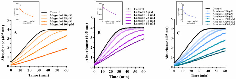
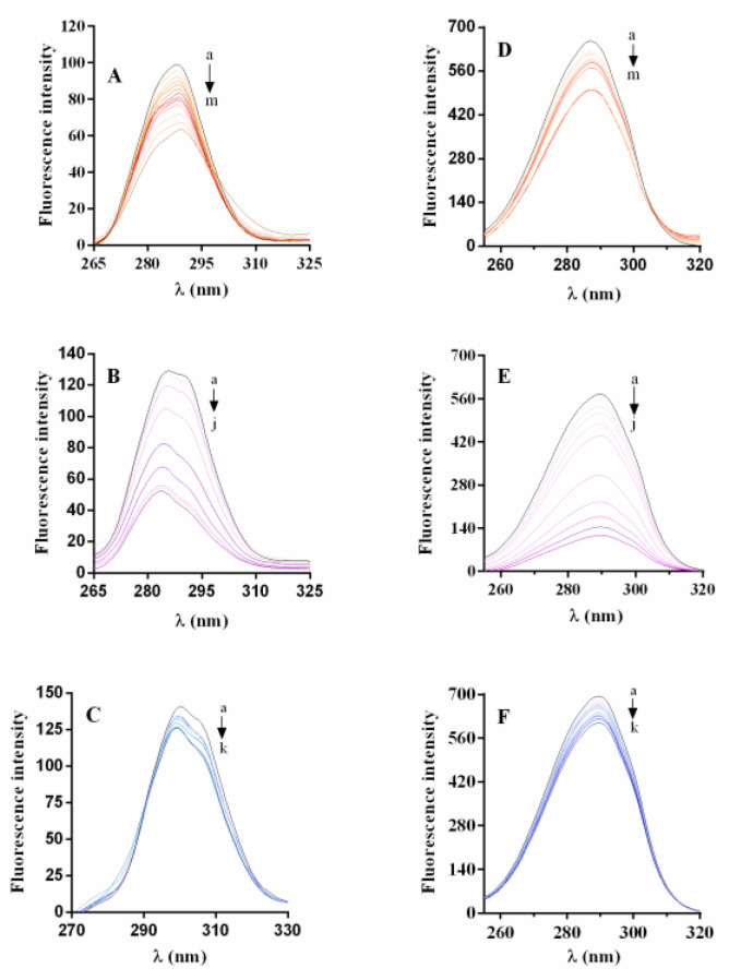
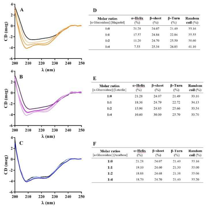
Magnolol and luteolin are two natural compounds recognized in several medicinal plants widely used in traditional medicine, including type 2 diabetes mellitus. This research aimed to determine the inhibitory activity of magnolol and luteolin on α-glucosidase activity. Their biological profile was studied by multispectroscopic methods along with inhibitory kinetic analysis and computational experiments. Magnolol and luteolin decreased the enzymatic activity in a concentration-dependent manner. With 0.075 µM α-glucosidase, the IC50 values were similar for both compounds (~ 32 µM) and significantly lower than for acarbose (815 μM). Magnolol showed a mixed-type antagonism, while luteolin showed a non-competitive inhibition mechanism. Thermodynamic parameters suggested that the binding of magnolol was predominantly sustained by hydrophobic interactions, while luteolin mainly exploited van der Waals contacts and hydrogen bonds. Synchronous fluorescence revealed that magnolol interacted with the target, influencing the microenvironment around tyrosine residues, and circular dichroism explained a rearrangement of the secondary structure of α-glucosidase from the initial α-helix to the final conformation enriched with β-sheet and random coil. Docking studies provided support for the experimental results. Altogether, the data propose magnolol, for the first time, as a potential α-glucosidase inhibitor and add further evidence to the inhibitory role of luteolin.
Keywords: circular dichroism; diabetes mellitus; enzymatic kinetics; hyperglycaemia; luteolin; magnolol; molecular docking; natural polyphenols; α-glucosidase inhibitors.
Conflict of interest statement
The authors declare no conflict of interest.
Figures


Similar articles
-
α-Glucosidase inhibition by luteolin: kinetics, interaction and molecular docking.Int J Biol Macromol. 2014 Mar;64:213-23. doi: 10.1016/j.ijbiomac.2013.12.007. Epub 2013 Dec 12. Int J Biol Macromol. 2014. PMID: 24333230
-
Inhibition of α-glucosidase by trilobatin and its mechanism: kinetics, interaction mechanism and molecular docking.Food Funct. 2022 Jan 24;13(2):857-866. doi: 10.1039/d1fo03636j. Food Funct. 2022. PMID: 34989743
-
Novel cinnamic acid magnolol derivatives as potent α-glucosidase and α-amylase inhibitors: Synthesis, in vitro and in silico studies.Bioorg Chem. 2021 Nov;116:105291. doi: 10.1016/j.bioorg.2021.105291. Epub 2021 Aug 19. Bioorg Chem. 2021. PMID: 34438122
-
Anti-α-Glucosidase and Antiglycation Activities of α-Mangostin and New Xanthenone Derivatives: Enzymatic Kinetics and Mechanistic Insights through In Vitro Studies.Molecules. 2022 Jan 15;27(2):547. doi: 10.3390/molecules27020547. Molecules. 2022. PMID: 35056861 Free PMC article.
-
Synthesis, in vitro inhibitory activity, kinetic study and molecular docking of novel N-alkyl-deoxynojirimycin derivatives as potential α-glucosidase inhibitors.J Enzyme Inhib Med Chem. 2020 Dec;35(1):1879-1890. doi: 10.1080/14756366.2020.1826941. J Enzyme Inhib Med Chem. 2020. PMID: 33003963 Free PMC article.
Cited by
-
Research Progress on Hypoglycemic Effects and Molecular Mechanisms of Flavonoids: A Review.Antioxidants (Basel). 2025 Mar 22;14(4):378. doi: 10.3390/antiox14040378. Antioxidants (Basel). 2025. PMID: 40298635 Free PMC article. Review.
-
Theoretical Studies for the Discovery of Potential Sucrase-Isomaltase Inhibitors from Maize Silk Phytochemicals: An Approach to Treatment of Type 2 Diabetes.Molecules. 2023 Sep 23;28(19):6778. doi: 10.3390/molecules28196778. Molecules. 2023. PMID: 37836621 Free PMC article.
-
Discovery of a Novel Chemo-Type for TAAR1 Agonism via Molecular Modeling.Molecules. 2024 Apr 11;29(8):1739. doi: 10.3390/molecules29081739. Molecules. 2024. PMID: 38675561 Free PMC article.
-
Comparative Evaluation of the Antiglycation and Anti-α-Glucosidase Activities of Baicalein, Baicalin (Baicalein 7-O-Glucuronide) and the Antidiabetic Drug Metformin.Pharmaceutics. 2022 Oct 9;14(10):2141. doi: 10.3390/pharmaceutics14102141. Pharmaceutics. 2022. PMID: 36297576 Free PMC article.
-
In Vitro Alpha-Glucosidase Inhibitory Activity and the Isolation of Luteolin from the Flower of Gymnanthemum amygdalinum (Delile) Sch. Bip ex Walp.Molecules. 2022 Mar 25;27(7):2132. doi: 10.3390/molecules27072132. Molecules. 2022. PMID: 35408529 Free PMC article.
References
Grants and funding
LinkOut - more resources
Full Text Sources
