. 2022 Feb 9;15(2):209.
doi: 10.3390/ph15020209.

A Lead-Based Fragment Library Screening of the Glycosyltransferase WaaG from Escherichia coli

Affiliations

A Lead-Based Fragment Library Screening of the Glycosyltransferase WaaG from Escherichia coli

Federico Riu et al. Pharmaceuticals (Basel). .

Abstract

Glucosyl transferase I (WaaG) in E. coli catalyzes the transfer of an α-d-glucosyl group to the inner core of the lipopolysaccharide (LPS) and plays an important role in the biogenesis of the outer membrane. If its activity could be inhibited, the integrity of the outer membrane would be compromised and the bacterium would be susceptible to antibiotics that are normally prevented from entering the cell. Herein, three libraries of molecules (A, B and C) were docked in the binding pocket of WaaG, utilizing the docking binding affinity as a filter to select fragment-based compounds for further investigations. From the results of the docking procedure, a selection of compounds was investigated by molecular dynamics (MD) simulations to obtain binding free energy (BFE) and KD values for ligands as an evaluation for the binding to WaaG. Derivatives of 1,3-thiazoles (A7 and A4) from library A and 1,3,4-thiadiazole (B33) from library B displayed a promising profile of BFE, with KD < mM, viz., 0.11, 0.62 and 0.04 mM, respectively. Further root-mean-square-deviation (RMSD), electrostatic/van der Waals contribution to the binding and H-bond interactions displayed a favorable profile for ligands A4 and B33. Mannose and/or heptose-containing disaccharides C1-C4, representing sub-structures of the inner core of the LPS, were also investigated by MD simulations, and compound C42- showed a calculated KD = 0.4 µM. In the presence of UDP-Glc2-, the best-docked pose of disaccharide C42- is proximate to the glucose-binding site of WaaG. A study of the variation in angle and distance was performed on the different portions of WaaG (N-, the C- domains and the hinge region). The Spearman correlation coefficient between the two variables was close to unity, where both variables increase in the same way, suggesting a conformational rearrangement of the protein during the MD simulation, revealing molecular motions of the enzyme that may be part of the catalytic cycle. Selected compounds were also analyzed by Saturation Transfer Difference (STD) NMR experiments. STD effects were notable for the 1,3-thiazole derivatives A4, A8 and A15 with the apo form of the protein as well as in the presence of UDP for A4.

Keywords: NMR spectroscopy; binding free energy; molecular docking; molecular dynamics.


Conflict of interest statement

The authors declare no conflict of interest.

Figures

Figure 1
Figure 1
Structural representation of outer (to the left) and inner (middle section) portions of the R1 core and the lipid A (to the right) of LPS [20]. R1 and R2 represent acyl groups attached to glucosamine residues of Lipid A. The structure is reported utilizing the Symbol Nomenclature For Glycans (SFNG) representation [21].
Figure 2
Figure 2
Structural formulas for rifampicin, chloramphenicol and colistin.
Scheme 1
Scheme 1
Flowchart of the computational and experimental study on WaaG and its interactions with compounds from libraries A, B and C.
Figure 3
Figure 3
Chemical backbone structure of the ligands from library A (a) and B (b); in the carbohydrate-based library C (c) R = HPO3Na. Figures S1–S5 report all ligands of libraries A, B and C.
Figure 4
Figure 4
Conformation of UDP-Glc2− in the binding pocket of WaaG, derived from U2F deposited co-crystallized structure (PDB ID: 2iw1).
Figure 5
Figure 5
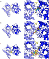
Docking poses of the representative ligands of libraries (a) A, (b) B and (c) C. On the right, a close-up of WaaG binding pocket showing the most relevant amino acids surrounding the ligands structures. (a) compound A1 in green, A7 in blue marine, A9 in yellow, A14 in orange and A16 in violet. (b) compound B3d in blue marine, B16 in light yellow, B22 in fuchsia, B26 in orange and B33 in green. (c) compound C6 in blue marine and C10 in yellow.
Figure 6
Figure 6
RMSD probability distributions obtained by a kernel density estimate using a normal distribution function. The calculation is done on the protein backbone of the apo-protein (from 2iw1) and the various WaaG-ligand complexes. The panels depict the RMSD distributions for WaaG in complex with ligands of (a) library A, (b) library B and (c) library C.
Figure 7
Figure 7
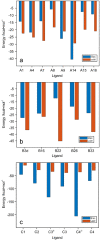
Contributions of electrostatic and van der Waals interactions to the binding of (a) library A, (b) library B and (c) library C ligands to WaaG binding pocket. Elec = electrostatic; vdW = van der Waals.
Figure 8
Figure 8
Hydrogen bond occupancies for ligands of (a) library A, (b) library B and (c) library C to the amino acids in the binding pocket of WaaG. Reported amino acids have at least 30% occupancy for one protein/ligand H-bond.
Figure 9
Figure 9
STD NMR spectra of ligand A4 and WaaG carried out with and without UDP as a competitor. In sequence from the bottom to the top, 1D 1H STD off-resonance (a) and on-resonance (b) irradiation of WaaG in the presence of A4 and off-resonance (c) and on-resonance (d) irradiation with UDP added to the protein-ligand mixture. Compound A4 shows interaction with WaaG also in the presence of UDP.
Figure 10
Figure 10
RMSD probability distributions obtained by a kernel density estimate using a normal distribution function. The calculation is done on the protein backbone of the apo-protein (from 2iw1, red) as a reference of RMSD values. Alignment of (a) the entire protein backbone, (b) N-domain, and (c) C-domain of apo, then RMSD calculation of the N- and C-domain and hinge region.
Figure 11
Figure 11
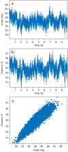
(a) Angle variation over time between the Cα atoms of His62 (N-domain), Lys248 (C-domain) and Gly168 (hinge region) and (b) distance fluctuations over time between the Cα atoms of His62 and Lys248 during the 10-ns MD calculation. (c) Scatter plot of the two variables against each other.
Figure 12
Figure 12
Graphical representation of the twist-like dynamics of WaaG. The C-domain of the protein (in cyan) is kept aligned, while the rest of the protein moves. Depicted are the two extremes (a,c) of the twist-like movements of the N-domain (in brick-red color) with respect to the C-domain and a representation of the two domains when aligned in the eclipsed state (b).
Figure 13
Figure 13
Molecular dynamics representation and interactions of ligands (a,b) A4 and (c,d) B33 with WaaG binding pocket. The structures are the most representative of the different clusters identified for each ligand.
Figure 14
Figure 14
(a) Molecular dynamics representation of C42− and (b,c) molecular docking conformation of C42− with WaaG-UDP-Glc2− complex.

References

    1. Croxen M.A., Finlay B.B. Molecular mechanisms of Escherichia coli pathogenicity. Nat. Rev. Microbiol. 2010;8:26–38. doi: 10.1038/nrmicro2265. - DOI - PubMed
    1. World Health Organization Prioritization of Pathogens to Guide Discovery, Research and Development of New Antibiotics for Drug Resistant Bacterial Infections, Including Tuberculosis. 2017. [(accessed on 14 January 2022)]. Available online: https://apps.who.int/iris/handle/10665/311820.
    1. Levin S. The crisis in antibiotic resistance. Infect. Dis. Clin. Pract. 1993;2:53. doi: 10.1097/00019048-199301000-00013. - DOI
    1. World Health Organization Outbreaks of E. coli O104:H4 Infection. 2011. [(accessed on 14 January 2022)]. Available online: https://www.euro.who.int/en/countries/germany/outbreaks-of-e.-coli-o104h....
    1. World Health Organization Antimicrobial Resistance. 2020. [(accessed on 14 January 2022)]. Available online: https://www.who.int/health-topics/antimicrobial-resistance.

LinkOut - more resources