. 2022 Feb 11;14(4):760.
doi: 10.3390/nu14040760.

Oligosaccharides Ameliorate Acute Kidney Injury by Alleviating Cluster of Differentiation 44-Mediated Immune Responses in Renal Tubular Cells

Affiliations

Oligosaccharides Ameliorate Acute Kidney Injury by Alleviating Cluster of Differentiation 44-Mediated Immune Responses in Renal Tubular Cells

Tso-Hsiao Chen et al. Nutrients. .

Abstract

Acute kidney injury (AKI) is a sudden episode of kidney damage that commonly occurs in patients admitted to hospitals. To date, no ideal treatment has been developed to reduce AKI severity. Oligo-fucoidan (FC) interferes with renal tubular cell surface protein cluster of differentiation 44 (CD44) to prevent renal interstitial fibrosis; however, the influence of oligosaccharides on AKI remains unknown. In this study, FC, galacto-oligosaccharide (GOS), and fructo-oligosaccharide (FOS) were selected to investigate the influence of oligosaccharides on AKI. All three oligosaccharides have been proven to be partially absorbed by the intestine. We found that the oligosaccharides dose-dependently reduced CD44 antigenicity and suppressed the hypoxia-induced expression of CD44, phospho-JNK, MCP-1, IL-1β, and TNF-α in NRK-52E renal tubular cells. Meanwhile, CD44 siRNA transfection and JNK inhibitor SP600125 reduced the hypoxia-induced expression of phospho-JNK and cytokines. The ligand of CD44, hyaluronan, counteracted the influence of oligosaccharides on CD44 and phospho-JNK. At 2 days post-surgery for ischemia-reperfusion injury, oligosaccharides reduced kidney inflammation, serum creatine, MCP-1, IL-1β, and TNF-α in AKI mice. At 7 days post-surgery, kidney recovery was promoted. These results indicate that FC, GOS, and FOS inhibit the hypoxia-induced CD44/JNK cascade and cytokines in renal tubular cells, thereby ameliorating AKI and kidney inflammation in AKI mice. Therefore, oligosaccharide supplementation is a potential healthcare strategy for patients with AKI.

Keywords: acute kidney injury; cluster of differentiation 44; fructo-oligosaccharide; galacto-oligosaccharide; inflammation; oligo-fucoidan.


Conflict of interest statement

The authors declare no conflict of interest.

Figures
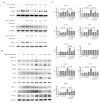
Figure 1
Figure 1
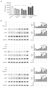
Inhibitory effect of oligosaccharides on CD44 signal transduction in hypoxic NRK-52E cells. The cells were pretreated with FC, FOS, or GOS for 30 min and then subjected to hypoxic culture. Protein expression was analyzed by Western blotting. (A) Dose-dependent effects of oligosaccharides on CD44 expression. (B) Effect of oligosaccharides on CD44-related signals. Oligosaccharide concentration was set as 0.1 mg/mL. Relative increases in protein bands are also presented in bar chart form (C). Results are expressed as mean ± SD (n = 4).
Figure 2
Figure 2
Role of CD44 and JNK in hypoxia-induced inflammatory responses in NRK-52E cells. Protein expression was analyzed by Western blot. (A) Inhibitory effect of CD44 siRNA transfection on phosphorylated JNK and cytokines. (B) Inhibitory effect of SP600125 on cytokines. The cells were pretreated with 20 μM SP600125 for 30 min prior to hypoxia treatment. Relative increases in protein bands are also presented in bar chart form. Results are expressed as mean ± SD (n = 4).
Figure 3
Figure 3
Promoting effect of cytokines secreted from hypoxic NRK-52E cells on the proliferation of macrophage RAW264.7. (A) Influence of oligosaccharides on the proliferation of RAW264.7 cells treated with 0.1 mg/mL FC, FOS, or GOS and cultured overnight. (B) Promoting effect of the culture medium of hypoxic NRK-52E cells on the proliferation of RAW264.7 cells. NRK-52E cells were pretreated with 0.1 mg/mL FC, FOS, or GOS for 30 min and then subjected to hypoxic culture. The culture medium of normal or hypoxic NRK-52E cells was taken out and used to culture RAW264.7 overnight. (C) MCP-1 level in the culture medium of hypoxic NRK-52E cells. (D) IL-1β level in the culture medium of hypoxic NRK-52E cells. Results are expressed as mean ± SD (n = 3).
Figure 4
Figure 4
Competitiveness of oligosaccharides and HA in binding to CD44. (A) Reducing effect of oligosaccharides on CD44 antigenicity. Recombinant rat CD44 (20 ng/mL) was mixed with or without FC, FOS, or GOS for 10 min and then analyzed using the CD44 ELISA kit. Results are expressed as mean ± SD (n = 4). *, p < 0.05 vs. the control group. (B) HA interfering with the inhibitory effect of oligosaccharides on CD44 and phosphorylated JNK expression. NRK-52E cells were pretreated with 0.1 mg/mL HA for 30 min, then administered with 0.1 mg/mL FC, FOS, or GOS for 30 min and finally subjected to hypoxic culture. Protein expression was analyzed by Western blot. Relative increases in the protein bands are also presented in bar chart form. Results are expressed as mean ± SD (n = 4).
Figure 5
Figure 5
Reducing effect of oligosaccharides on serum creatinine and cytokines in AKI mice at the early stage. Blood was collected from each mouse 2 days after IRI surgery to measure serum creatinine (A), MCP-1 (B), IL-1β (C), and TNF-α (D). The results are expressed as means ± SD (n = 8).
Figure 6
Figure 6
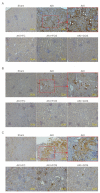
Inhibitory effect of oligosaccharides on renal inflammation in mice with early-stage AKI. The kidneys from each mouse 2 days after IRI surgery were collected for IHC staining. (A) Ly6G IHC staining. The white arrow indicates Ly6G positive staining neutrophils. (B) F4/80 IHC staining. The white arrow indicates F4/80 positive staining macrophages. (C) TNF-α IHC staining.
Figure 7
Figure 7
Reducing effect of oligosaccharides on the severity of AKI in post-AKI. Blood and kidneys from each mouse were collected 7 days after IRI surgery for biochemical analysis and IHC staining, respectively. (A) Serum creatinine levels. The results are expressed as means ± SD (n = 8). n.s., no significance. (B) Serum NGAL levels. The results are expressed as means ± SD (n = 8). (C) PAS staining of mouse kidney cortex. Pink staining of brush border is visible in health renal tubules, and loss of brush border is evident in the unhealthy dilated tubules.

Similar articles

Cited by

References

    1. Makris K., Spanou L. Acute Kidney Injury: Definition, Pathophysiology and Clinical Phenotypes. Clin. Biochem. Rev. 2016;37:85–98. - PMC - PubMed
    1. Silver S.A., Harel Z., McArthur E., Nash D.M., Acedillo R., Kitchlu A., Garg A.X., Chertow G.M., Bell C.M., Wald R. Causes of Death after a Hospitalization with AKI. J. Am. Soc. Nephrol. 2018;29:1001–1010. doi: 10.1681/ASN.2017080882. - DOI - PMC - PubMed
    1. Joannidis M., Druml W., Forni L.G., Groeneveld A.B.J., Honore P.M., Hoste E., Ostermann M., Oudemans-van Straaten H.M., Schetz M. Prevention of acute kidney injury and protection of renal function in the intensive care unit: Update 2017: Expert opinion of the Working Group on Prevention, AKI section, European Society of Intensive Care Medicine. Intensive Care Med. 2017;43:730–749. doi: 10.1007/s00134-017-4832-y. - DOI - PMC - PubMed
    1. Bijuklic K., Jennings P., Kountchev J., Hasslacher J., Aydin S., Sturn D., Pfaller W., Patsch J.R., Joannidis M. Migration of leukocytes across an endothelium-epithelium bilayer as a model of renal interstitial inflammation. Am. J. Physiol. Physiol. 2007;293:C486–C492. doi: 10.1152/ajpcell.00419.2006. - DOI - PubMed
    1. Bihorac A., Baslanti T.O., Cuenca A.G., Hobson C., Ang D., Efron P.A., Maier R.V., Moore F.A., Moldawer L.L. Acute kidney injury is associated with early cytokine changes after trauma. J. Trauma Acute Care Surg. 2013;74:1005–1013. doi: 10.1097/TA.0b013e31828586ec. - DOI - PMC - PubMed

Substances