. 2022 Feb 13;14(4):786.
doi: 10.3390/nu14040786.

Cancer Cell Inhibiting Sea Cucumber (Holothuria leucospilota) Protein as a Novel Anti-Cancer Drug

Affiliations

Cancer Cell Inhibiting Sea Cucumber (Holothuria leucospilota) Protein as a Novel Anti-Cancer Drug

Ruizhen Ru et al. Nutrients. .

Abstract

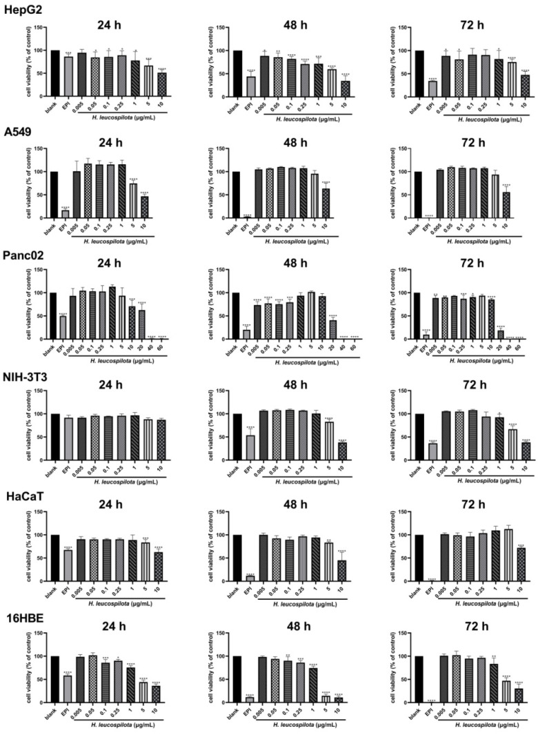
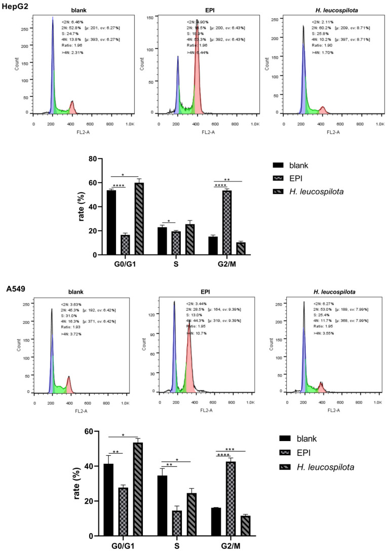
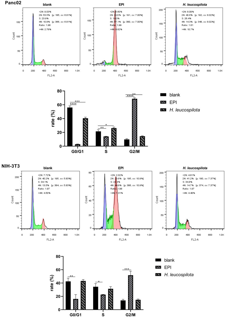
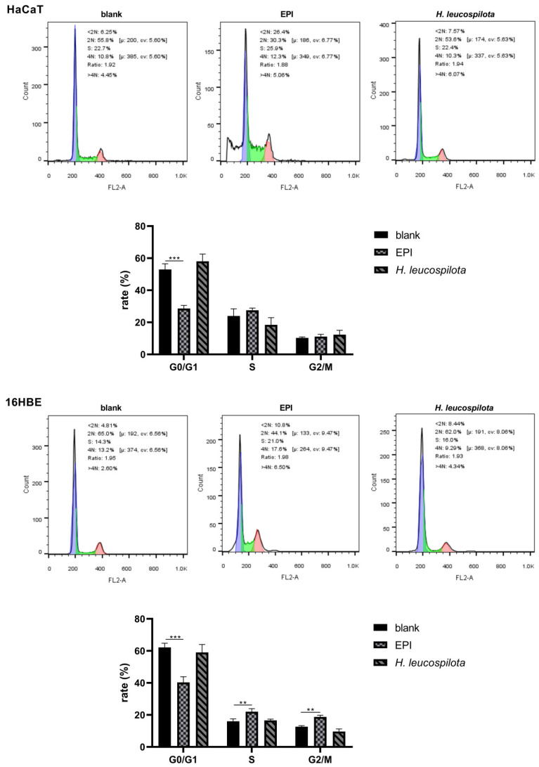
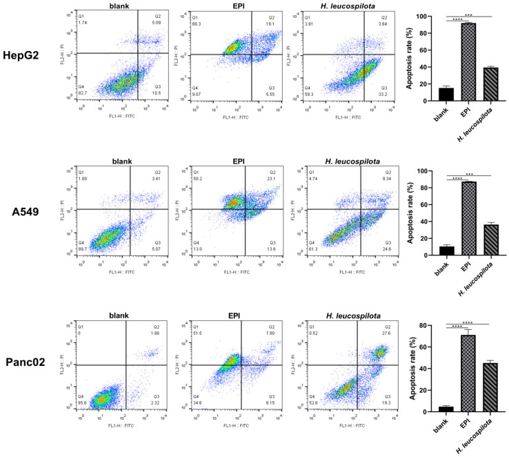
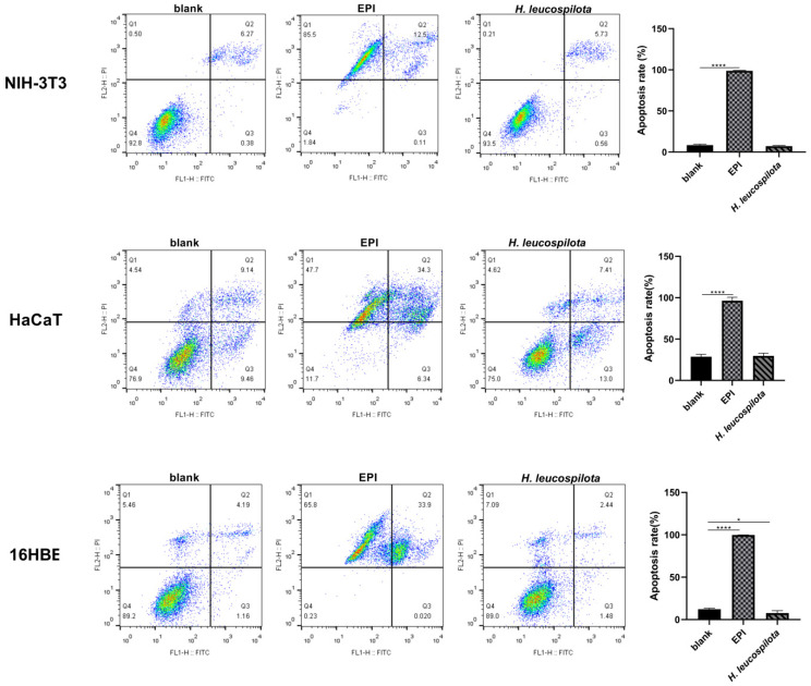
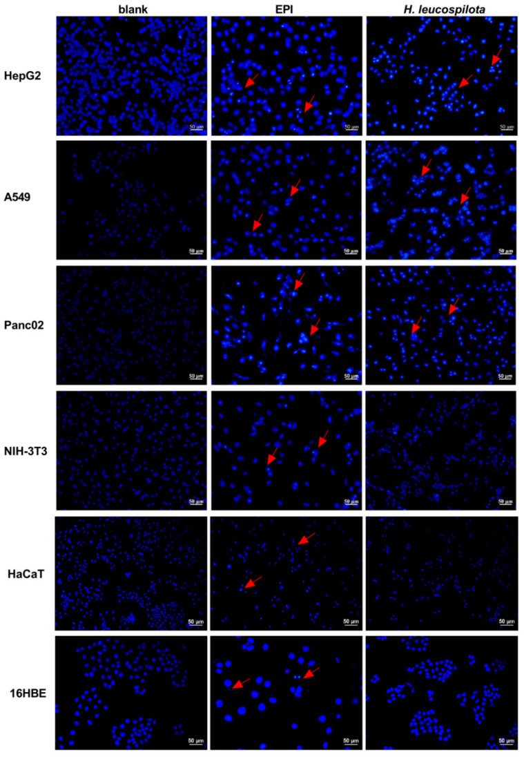
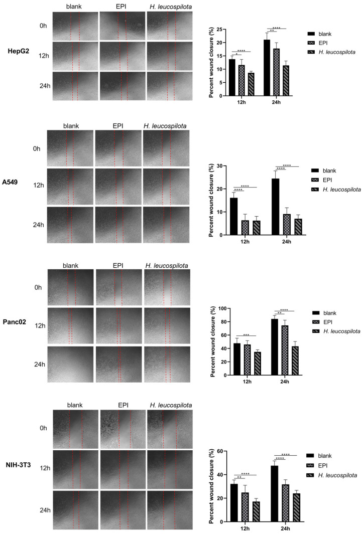
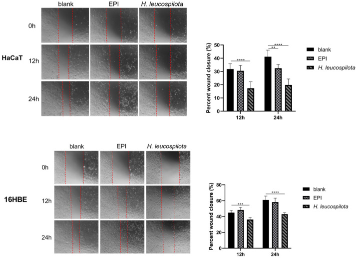
Cancer remains the primary cause of death worldwide. To develop less toxic anti-cancer drugs to relieve the suffering and improve the survival of cancer patients is the major focus in the anti-cancer field. To this end, marine creatures are being extensively studied for their anti-cancer effects, since extracts from at least 10% of the marine organisms have been shown to possess anti-tumor activities. As a classic Chinese traditional medicine, sea cucumbers and compounds extracted from the sea cucumbers, such as polysaccharides and saponins, have recently been shown to exhibit anti-cancer, anti-inflammatory, and anti-oxidant effects. Holothuria leucospilota (H. leucospilota) is a tropical edible sea cucumber species that has been successfully cultivated and farmed in large scales, providing a readily available source of raw materials to support the development of novel marine anti-cancer drugs. However, very few studies have so far been performed on the biological activities of H. leucospilota. In this study, we first investigated the anti-cancer effect of H. leucospilota protein on three cancer cell lines (i.e., HepG2, A549, Panc02) and three normal cell lines (NIH-3T3, HaCaT, 16HBE). Our data showed that H. leucospilota protein decreased the cell viabilities of HepG2, A549, HaCaT, 16HBE in a concentration-dependent manner, while Panc02 and NIH-3T3 in a time- and concentration-dependent manner. We also found that the inhibitory effect of H. leucospilota protein (≥10 μg/mL) on cell viability is near or even superior to EPI, a clinical chemotherapeutic agent. In addition, our data also demonstrated that H. leucospilota protein significantly affected the cell cycle and induced apoptosis in the three cancer cell lines investigated; in comparison, it showed no effects on the normal cell lines (i.e., NIH-3T3, HaCaT and 16HBE). Finally, our results also showed that H. leucospilota protein exhibited the excellent performance in inhibiting cell immigrations. In conclusion, H. leucospilota protein targeted the cancer cell cycles and induced cancer cell apoptosis; its superiority to inhibit cancer cell migration compared with EPI, shows the potential as a promising anti-cancer drug.

Keywords: anti-cancer activity; cell apoptosis; cell migration; marine extractions; sea cucumber protein; targeted effects.


Conflict of interest statement

There are no conflict of interest to declare.

Figures

Figure 1
Figure 1
Inhibition of cellular viability by Holothuria leucospilota (H. leucospilota) protein. Exponentially growing HepG2, A549, Panc02, NIH-3T3, HaCaT and 16HBE cells were treated with EPI (10 μΜ) and the increased concentrations of H. leucospilota protein for 24, 48, 72 h. Viable cells were assayed as described in Materials and Methods. All experiments were repeated as least three times. Data are expressed as Mean ± SD. * p < 0.05, ** p < 0.01, *** p < 0.001, **** p < 0.0001.
Figure 2
Figure 2
Effect of H. leucospilota protein on cell cycle distribution. Cells were treated with the IC50 concentrations of H. leucospilota protein for 48 h, with EPI (10 μΜ) as positive control. All experiments were repeated at least three times. Data are mean ± SD. * p < 0.05, ** p < 0.01, *** p < 0.001, **** p < 0.0001.
Figure 2
Figure 2
Effect of H. leucospilota protein on cell cycle distribution. Cells were treated with the IC50 concentrations of H. leucospilota protein for 48 h, with EPI (10 μΜ) as positive control. All experiments were repeated at least three times. Data are mean ± SD. * p < 0.05, ** p < 0.01, *** p < 0.001, **** p < 0.0001.
Figure 3
Figure 3
Effect of H. leucospilota protein treatment on cell apoptosis. Cells were treated with the IC50 concentrations of H. leucospilota protein for 48 h, and EPI (10 μΜ) as positive group. * p < 0.05, *** p < 0.001, **** p < 0.0001.
Figure 3
Figure 3
Effect of H. leucospilota protein treatment on cell apoptosis. Cells were treated with the IC50 concentrations of H. leucospilota protein for 48 h, and EPI (10 μΜ) as positive group. * p < 0.05, *** p < 0.001, **** p < 0.0001.
Figure 4
Figure 4
Effect of H. leucospilota protein treatment on cell morphology. Cells were treated with the IC50 concentrations of H. leucospilota protein for 48 h, and EPI (10 μΜ) as positive group. The morphological characteristics of apoptosis were shrunken, hyperchromatic, and pyknotic cells.
Figure 5
Figure 5
Effect of H. leucospilota protein treatment on cellular migration. Wounds were introduced in HepG2, A549, Panc02, NIH-3T3, HaCaT and 16HBE confluent mono-layers cultured in the presence or absence (control) with the IC50 concentrations of H. leucospilota protein for 0, 12, and 24 h, and EPI (10 μΜ) as positive group. Migration rate of HepG2, A549, Panc02, NIH-3T3, HaCaT, 16HBE in Figure. Experiments were repeated at least three times. Data are expressed as mean ± SD. * p < 0.05, ** p < 0.01, *** p < 0.001, **** p < 0.0001.
Figure 5
Figure 5
Effect of H. leucospilota protein treatment on cellular migration. Wounds were introduced in HepG2, A549, Panc02, NIH-3T3, HaCaT and 16HBE confluent mono-layers cultured in the presence or absence (control) with the IC50 concentrations of H. leucospilota protein for 0, 12, and 24 h, and EPI (10 μΜ) as positive group. Migration rate of HepG2, A549, Panc02, NIH-3T3, HaCaT, 16HBE in Figure. Experiments were repeated at least three times. Data are expressed as mean ± SD. * p < 0.05, ** p < 0.01, *** p < 0.001, **** p < 0.0001.

Similar articles

Cited by

References

    1. Siegel R.L., Kimberly D.M., Hannah E.F., Ahmedin J. Cancer Statistics, 2021. CA Cancer J. Clin. 2021;71:7–33. doi: 10.3322/caac.21654. - DOI - PubMed
    1. Sung H., Ferlay J., Siegel R.L., Laversanne M., Soerjomataram I., Jemal A., Bray F. Global Cancer Statistics 2020: GLOBOCAN Estimates of Incidence and Mortality Worldwide for 36 Cancers in 185 Countries. CA Cancer J. Clin. 2021;71:209–249. doi: 10.3322/caac.21660. - DOI - PubMed
    1. Yang Y.-M., Hong P., Xu W.W., He Q.-Y., Li B. Advances in targeted therapy for esophageal cancer. Signal Transduct. Target. Ther. 2020;5:229. doi: 10.1038/s41392-020-00323-3. - DOI - PMC - PubMed
    1. Hu J., Li Y., Li H., Shi F., Xie L., Zhao L., Tang M., Luo X., Jia W., Fan J., et al. Targeting Epstein-Barr virus oncoprotein LMP1-mediated high oxidative stress suppresses EBV lytic reactivation and sensitizes tumors to radiation therapy. Theranostics. 2020;10:11921–11937. doi: 10.7150/thno.46006. - DOI - PMC - PubMed
    1. Kong F.F., Ying C.R., Kong F., Ying H., Du C., Huang S., Zhou J., Chen J., Sun L., Chen X., et al. Patterns of local-regional failure after primary intensity modulated radiotherapy for nasopharyngeal carcinoma. Radiat. Oncol. 2014;9:60. doi: 10.1186/1748-717X-9-60. - DOI - PMC - PubMed