Review
. 2022 Jan 19;14(2):188.
doi: 10.3390/v14020188.

RNA-Binding Proteins as Regulators of Internal Initiation of Viral mRNA Translation

Affiliations
Review

RNA-Binding Proteins as Regulators of Internal Initiation of Viral mRNA Translation

Brenda López-Ulloa et al. Viruses. .

Abstract

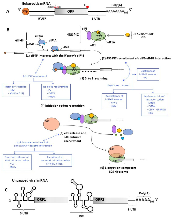
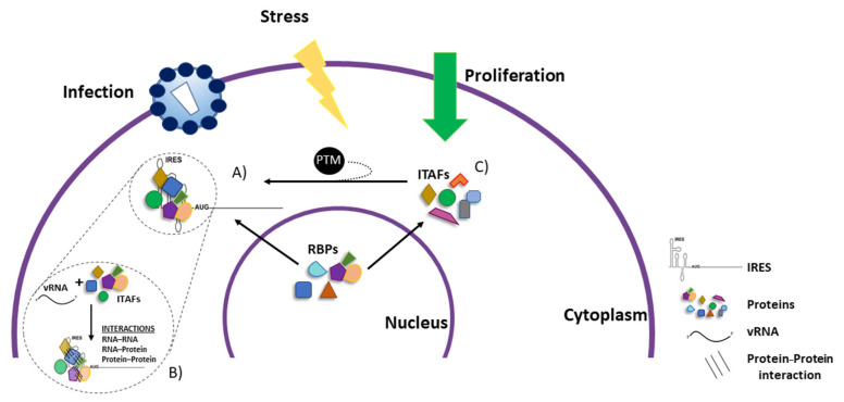
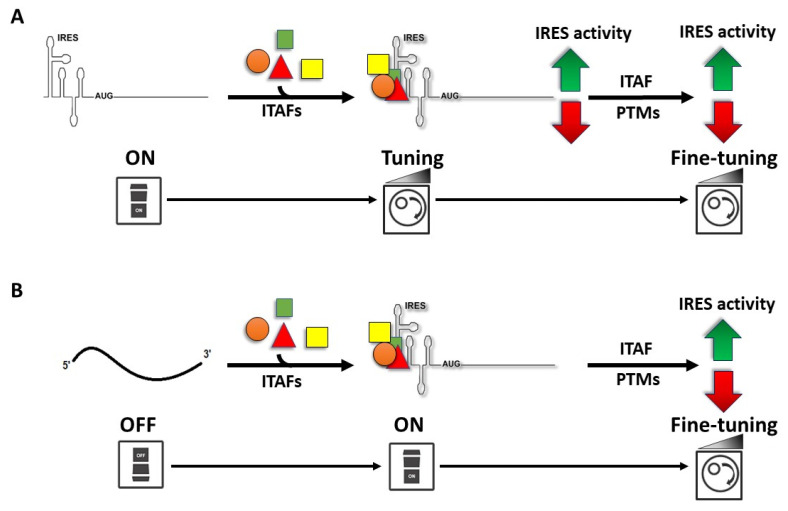
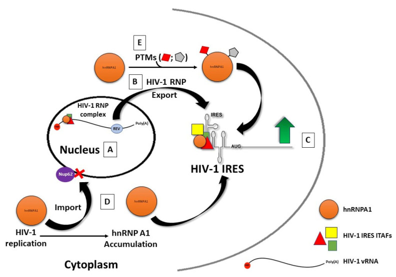
Viruses are obligate intracellular parasites that depend on the host's protein synthesis machinery for translating their mRNAs. The viral mRNA (vRNA) competes with the host mRNA to recruit the translational machinery, including ribosomes, tRNAs, and the limited eukaryotic translation initiation factor (eIFs) pool. Many viruses utilize non-canonical strategies such as targeting host eIFs and RNA elements known as internal ribosome entry sites (IRESs) to reprogram cellular gene expression, ensuring preferential translation of vRNAs. In this review, we discuss vRNA IRES-mediated translation initiation, highlighting the role of RNA-binding proteins (RBPs), other than the canonical translation initiation factors, in regulating their activity.

Keywords: IRES; IRES-transacting factor; ITAF; RBP; RNA-binding protein; internal ribosome entry site.


Conflict of interest statement

The authors declare no conflict of interest.

Figures

Figure 1
Figure 1
Cap-dependent translation initiation and viral strategies for ribosomal recruitment. (A) Schematic representation of a eukaryotic mRNA, including the 5′ cap structure, the 5′ untranslated region (UTR), the open reading frame (ORF), followed by the 3′ UTR and the Poly(A) at the 3′ end. The initiation codon (red) is also highlighted as part of the optimal context. (B) Schematic representation of canonical cap-dependent translation initiation summarized in 6 steps. Circularization of the mRNA is omitted. The eIF4F complex, 43S-preinitiation complex (PIC), and ternary complex (TC) are shown. The presence of the cap structure at the 5′ end of the mRNA allows for the recruitment of the eIF4F complex (1) and the 43S PIC (2). After these complexes scan the mRNA, they travel in an ATP-dependent, 5′ to 3′ direction (3). During scanning, eIF4A, in association with cofactor eIF4B, unwinds the secondary structure of the 5′ UTR. Scanning ends when the initiation codon is recognized (4). The factor eIF1 and eIF1A help avoid non-initiation codon interaction with the tRNAMeti placed in the P-site of the 40S subunit. Codon:anticodon interaction allows for the hydrolysis of eIF2-GTP and triggers the release of several factors, contributing to the recruitment of the 60S subunit (5) and the release of the remaining initiation factors, leading to the assembly of an elongation-competent 80S ribosome (6). Some viral strategies to recruit the ribosome by IRES are shown in blue: (a) whether intact eIF4F complex is needed; (b) regarding the position of the 40S subunit recruitment; and (c) whether the vRNA directly interacts with the ribosome. (C) Schematic representation of Dicistroviridae RNA, highlighting the IRES present in the 5′ UTR and the intergenic region (IGR).
Figure 2
Figure 2
Nuclear and Cytoplasmic RBPs act as ITAFs. (A,B) IRES-transacting factors (ITAFs) are cytoplasmic or nucleus–cytoplasmic shuttling RNA-binding proteins (RBPs) [82,222]. (C) These ITAF–RNA and ITAF–ITAF interactions depend on cellular conditions such as stress, proliferation, or viral infection, which may trigger specific ITAF PTMs, that reduce or enhance the RBPs affinity for its target RNA. (D) ITAFs assist in RNA structuring by establishing multiple contacts with the same RNA molecules and other RBP [82,95,96,147,168,223]. Thus, RBPs induce RNA conformations or RNP complexes with differential affinities for the translation machinery.
Figure 3
Figure 3
Model of IRES translational control mediated by RNA protein binding. (A) Some vRNAs do not require ITAFs to assemble (ON state). However, the association of different RBPs regulates the rate of translation initiation. ITAFs can also suffer PTMs, impacting their function over the IRES, further stimulating (green arrow) or reducing (red arrow) its activity [225]. (B) Some vRNAs (OFF state) require RBPs to assemble a functional IRES (ON state) [68,198]. ITAF PTMs can impact the ITAF localization, RNA affinity, or affinity for other RBPs impacting IRES function [81,225].
Figure 4
Figure 4
HnRNPA1: a model ITAF for the HIV-1 IRES. (A) In HIV-1 replicating cells, hnRNPA1 associates with the HIV-1 vRNA in the nucleus as part of the HIV-1 RNP [212,367]. (B) The HIV-1 RNP export is mediated by the viral protein Rev (reviewed in [367,373,374]). (C) HnRNPA1 stimulates (green arrow) HIV-1 IRES-mediated translation initiation [212,225]. (D) In cells, HIV-1 replication increases hnRNPA1 expression [212]. Cytoplasmic hnRNP A1 is re-imported into the nucleus by bound Trn-1, which interacts with the core member of the nuclear pore complex Nup62. However, in cells replicating HIV-1, Nup62 is downregulated, leading to the cytoplasmic retention of hnRNPA1. Cytoplasmic accumulation of hnRNPA1 further increases HIV-1 IRES activity [212]. (E) Post-translational modifications (PTMs) of hnRNPA1, including Mnk-mediated phosphorylation and PRMT5-induced symmetrical di-methylation of arginine residues, further promote HIV-1 IRES activity [212,225].

Similar articles

Cited by

References

    1. Abrahao J., Silva L., Silva L.S., Khalil J.Y.B., Rodrigues R., Arantes T., Assis F., Boratto P., Andrade M., Kroon E.G., et al. Tailed giant Tupanvirus possesses the most complete translational apparatus of the known virosphere. Nat. Commun. 2018;9:749. doi: 10.1038/s41467-018-03168-1. - DOI - PMC - PubMed
    1. Stern-Ginossar N., Thompson S.R., Mathews M.B., Mohr I. Translational Control in Virus-Infected Cells. Cold Spring Harb. Perspect. Biol. 2019;11:a033001. doi: 10.1101/cshperspect.a033001. - DOI - PMC - PubMed
    1. Jan E., Mohr I., Walsh D. A Cap-to-Tail Guide to mRNA Translation Strategies in Virus-Infected Cells. Annu. Rev. Virol. 2016;3:283–307. doi: 10.1146/annurev-virology-100114-055014. - DOI - PubMed
    1. Walsh D., Mohr I. Viral subversion of the host protein synthesis machinery. Nat. Rev. Microbiol. 2011;9:860–875. doi: 10.1038/nrmicro2655. - DOI - PMC - PubMed
    1. Schneider R.J., Mohr I. Translation initiation and viral tricks. Trends Biochem. Sci. 2003;28:130–136. doi: 10.1016/S0968-0004(03)00029-X. - DOI - PubMed

Publication types