. 2022 Jan 20;14(2):201.
doi: 10.3390/v14020201.

SARS-CoV-2: Ultrastructural Characterization of Morphogenesis in an In Vitro System

Affiliations

SARS-CoV-2: Ultrastructural Characterization of Morphogenesis in an In Vitro System

Debora Ferreira Barreto-Vieira et al. Viruses. .

Abstract

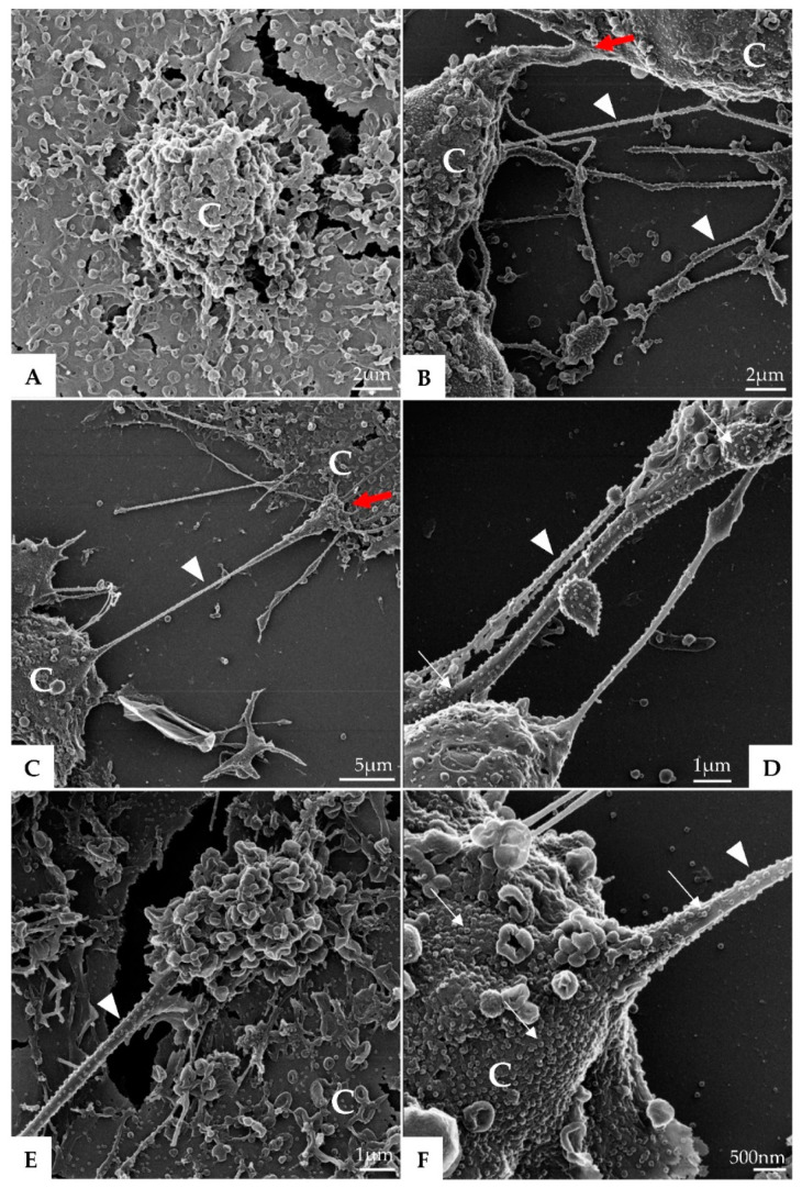
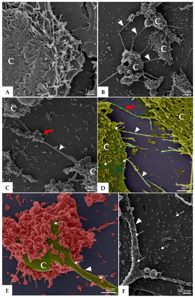
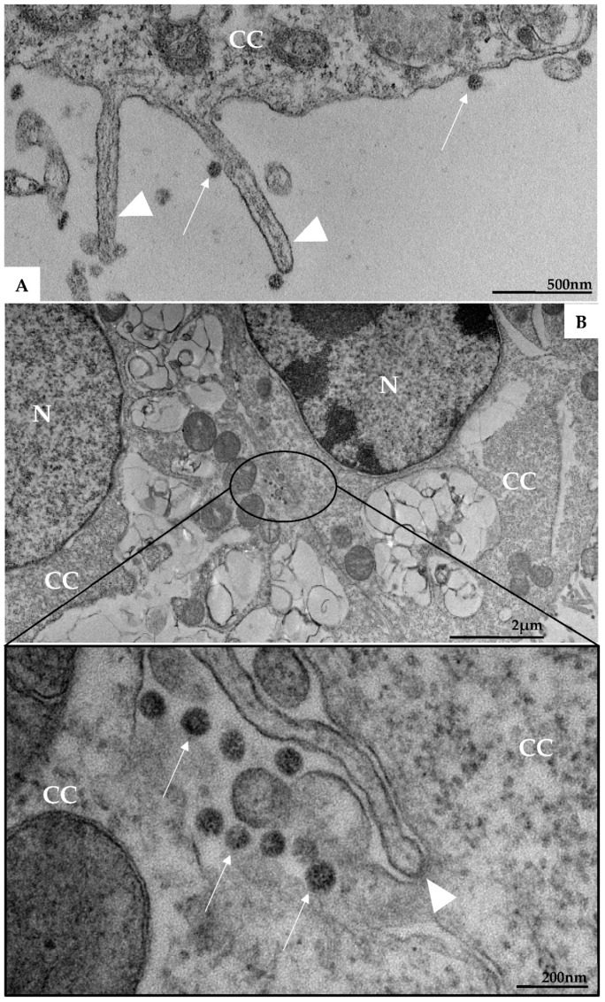
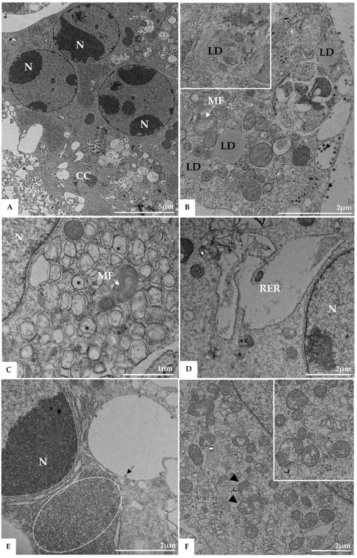
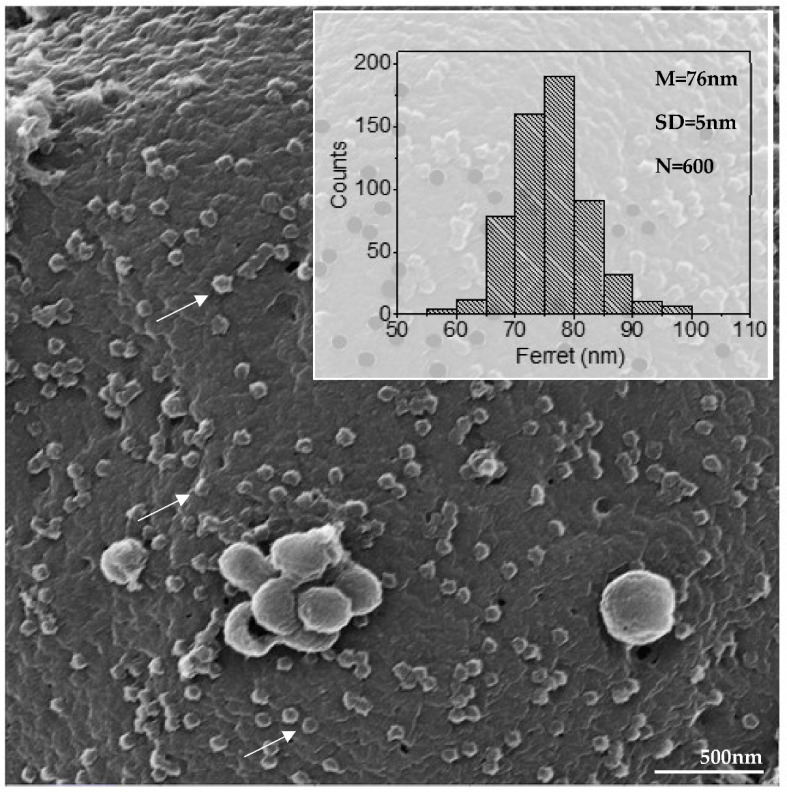
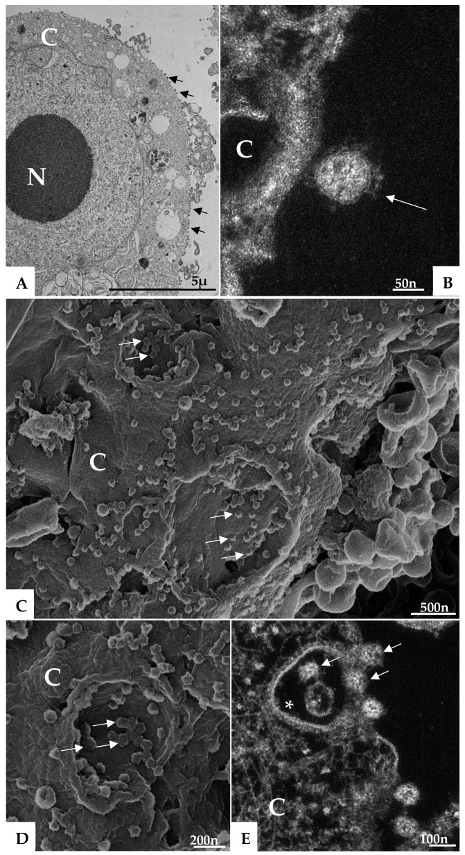
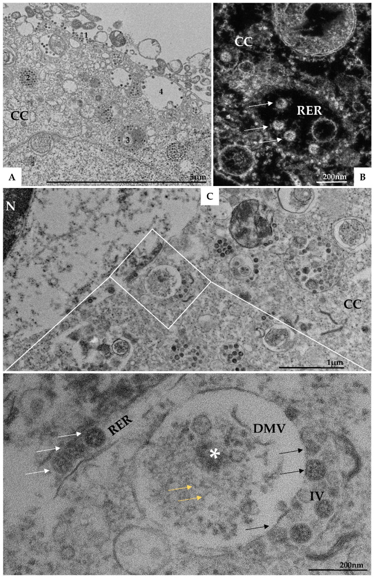
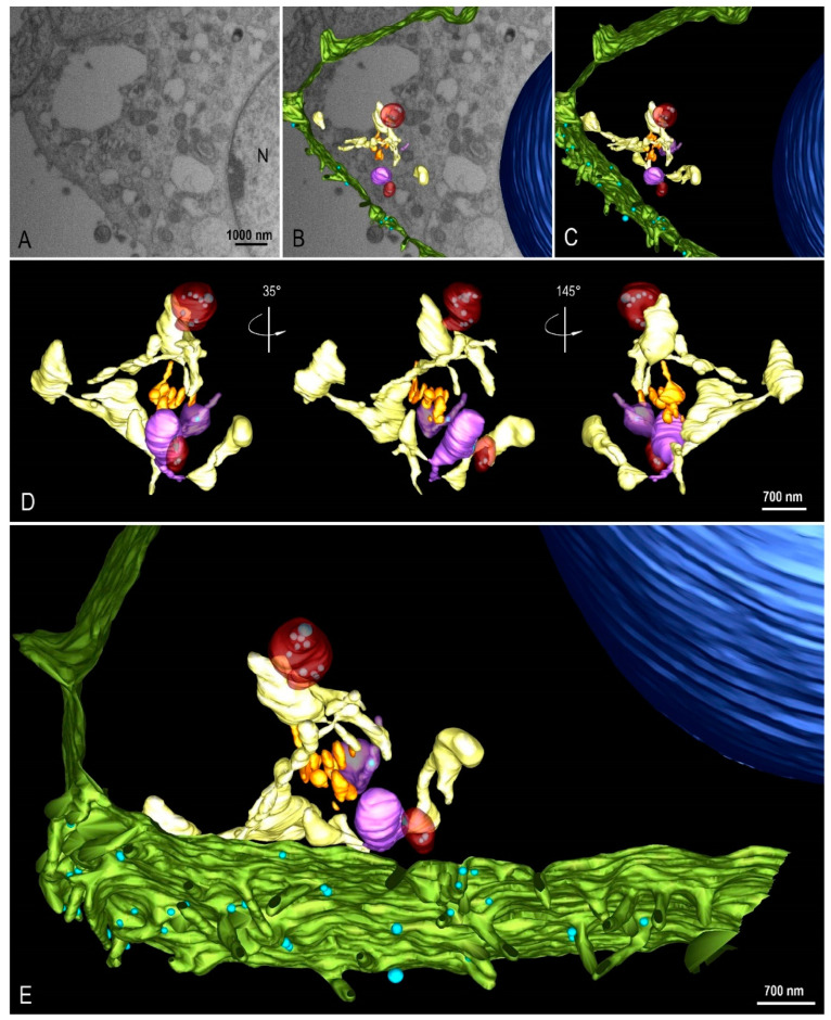
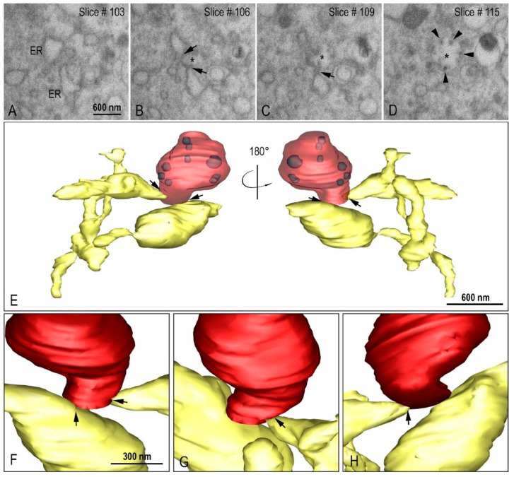
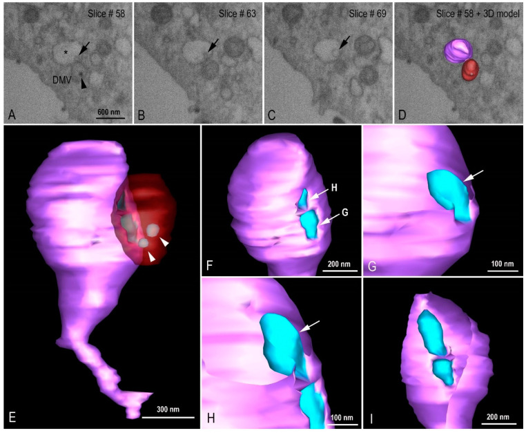
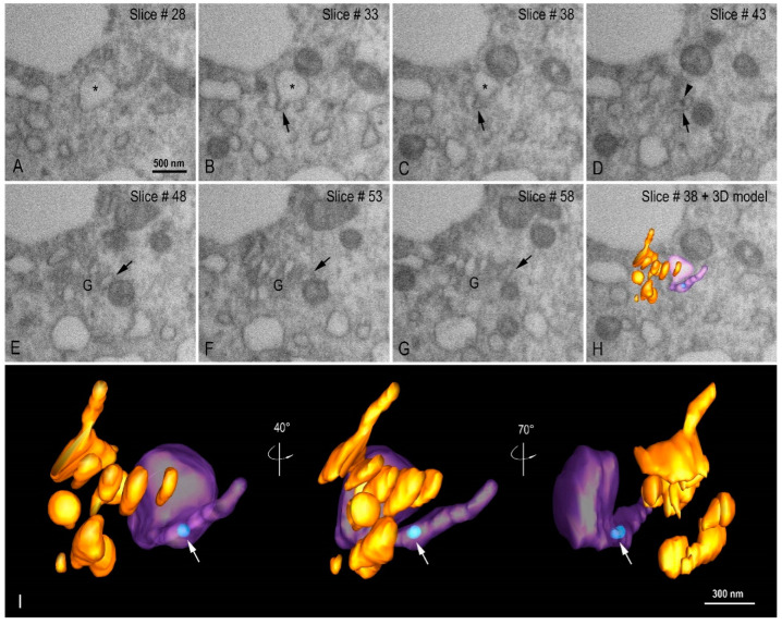
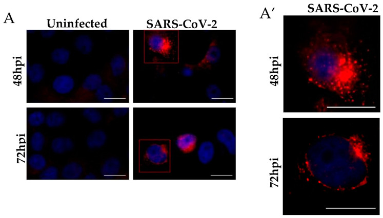
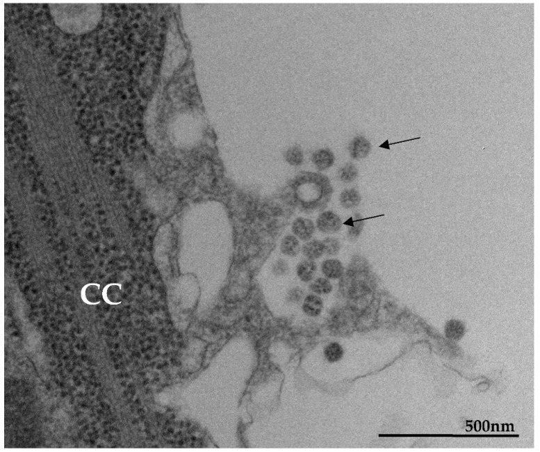
The pandemic caused by the severe acute respiratory syndrome coronavirus 2 (SARS-CoV-2) has impacted public health and the world economy and fueled a worldwide race to approve therapeutic and prophylactic agents, but so far there are no specific antiviral drugs. Understanding the biology of the virus is the first step in structuring strategies to combat it, and in this context several studies have been conducted with the aim of understanding the replication mechanism of SARS-CoV-2 in vitro systems. In this work, studies using transmission and scanning electron microscopy and 3D electron microscopy modeling were performed with the goal of characterizing the morphogenesis of SARS-CoV-2 in Vero-E6 cells. Several ultrastructural changes were observed-such as syncytia formation, cytoplasmic membrane projections, lipid droplets accumulation, proliferation of double-membrane vesicles derived from the rough endoplasmic reticulum, and alteration of mitochondria. The entry of the virus into cells occurred through endocytosis. Viral particles were observed attached to the cell membrane and in various cellular compartments, and extrusion of viral progeny took place by exocytosis. These findings allow us to infer that Vero-E6 cells are highly susceptible to SARS-CoV-2 infection as described in the literature and their replication cycle is similar to that described with SARS-CoV and MERS-CoV in vitro models.

Keywords: 3D electron microscopy modeling; SARS-CoV-2; Vero-E6 cells; morphogenesis; scanning electron microscopy; transmission electron microscopy; ultrastructural studies.


Conflict of interest statement

The authors declare no conflict of interest exists.

Figures

Figure 1
Figure 1
Vero-E6 cells 48 h post-infection with SARS-CoV-2 (HIM images). (A) Uninfected Vero-E6 cell at 48 h of cultivation (cell control). (BF) Infected cells presenting filopodia (arrowhead); connection (red arrow) between adjacent cells mediated by filopodia was observed. Virus particles (thick arrow) attached to cell filopodia and with cell membrane were observed. Cell (C).
Figure 2
Figure 2
Vero-E6 cells 72 h post-infection with SARS-CoV-2 (HIM images). (A) Uninfected Vero-E6 cell at 72 h of cultivation (cell control). (BE) Infected Vero-E6 monolayer, connection (red arrow) between cells mediated by filopodia was observed (BD). Virus particles (thick arrow, blue structures) was detected attached to cell filopodia (green, image (D)) and with cell membrane (green or red, image (DF). Cell (C), filopodia (arrowhead). The images (D,E) were colored in Adobe photoshop.
Figure 3
Figure 3
Microvilli in Vero-E6 cells 48 h post-infection with SARS-CoV-2 (TEM images). Microvilli (arrowhead) presenting adsorbed SARS-CoV-2 particles (thick arrow) (A). Interaction between adjacent cells mediated by Microvilli (B). Microvilli (arrowhead), cell cytoplasm (CC), nucleus (N).
Figure 4
Figure 4
Syncytia formation (A), proliferation of lipid droplets (LD) (B), and double membrane vesicle (*) (C), myelin figures (MF) (B,C), thickening of the rough endoplasmic reticulum (RER) (D), electron-dense ribosomes (marked area) (E), and degeneration of mitochondria (arrowhead) (F) in Vero-E6 cells 48 h post-infection with SARS-CoV-2 (TEM images). Virus particles (arrow), cell cytoplasm (CC), nucleus (N).
Figure 5
Figure 5
Diameter of SARS-CoV-2 particles attached in cell membrane (arrow) (HIM image). The average diameter of the SARS-CoV-2 particles was around 76 nanometers. Mean (M), standard deviation (SD), number of measured particles (N), virus particles (arrow).
Figure 6
Figure 6
Attachment and endocytosis of SARS-CoV-2 particles (TEM (A), HAADF-STEM (B,E) and HIM (C,D) images). SARS-CoV-2 particles (arrows) attached to the cell membrane (AD) and being internalized by endocytosis (arrow) (E). Endocytic vesicles coated in clathrin (*) (E). Cell cytoplasm (CC), cell membrane (CM), nucleus (N).
Figure 7
Figure 7
SARS-CoV-2 particles in lumen of several cellular compartments. (A) SARS-CoV-2 particles attached to the cell membrane (1), on endosomes (2), on the rough endoplasmic reticulum (3 and image (B) (arrow)), and being exocytosed via vesicles (4). (C) Presence of double membrane vesicle (DMV) containing in their lumen electron-dense material (*) and RNA-like filaments (yellow arrow). Budding of viral particles (black arrow) from these vesicles to into intermediate vesicles (IV) was observed. Virions were observed in rough endoplasmic reticulum cisterns (white arrow). Cell cytoplasm (CC), nucleus (N), and rough endoplasmic reticulum cisterns (RER). Transmission electron microscopy.
Figure 8
Figure 8
3D modeling of Vero-E6 48 h post-infection with SARS-CoV-2. (A) FIB-SEM image of the block surface showing an infected cell. (B,C) Combination of a block surface image and the 3D model of structures of interest. (D) Different angle views of a region of interest showing the organization and interaction between different cellular compartments. (E) 3D model showing a global view of the model. Green (cell membrane), light blue (virus particle), dark blue (nucleus), yellow (rough endoplasmic reticulum), orange (Golgi complex), red (double membrane vesicle), purple (intermediate vesicle). See video in Supplementary Material (Video S1).
Figure 9
Figure 9
Interaction between the rough endoplasmic reticulum (ER) and double membrane vesicle (DMV). (AD) FIB-SEM image sequence showing the ER and its connection (arrows) to a DMV (asterisk). Some virus particles can be seen inside the DMV (arrow heads). (EH) 3D model of the DMV (red), virus particles (purple) inside the DMV and the ER (yellow). The regions of connection of the ER to the DMV are indicated with arrows. Interconnections within the ER network can be observed.
Figure 10
Figure 10
Virion budding events at intermediate vesicle (IV). (AD) FIB-SEM image sequence exhibiting different virus budding stages (arrows) at IV (asterisk) and the proximity with a double membrane vesicle (DMV). Virus particles can be observed inside the DMV (arrow head). (EI) 3D model showing the DMV (red with transparence), virus particles (light blue, arrow heads), and the IV (purple). (F) Extra vesicular view of two virus particle budding regions. The arrows indicate the cutting plane showed in G and H. (G) Initial stage of virus particle budding. (H) Final stage of virus particle budding. (I) Intra vesicular view of the virus budding. See video in Supplementary Material (Video S2).
Figure 11
Figure 11
Organization of the Golgi complex (G) and intermediate vesicle (IV). (AH) FIB-SEM image sequence showing an IV (asterisk) with a tubular region (arrow) in close proximity with to the Golgi complex. A virus particle can be seen inside the tubular region of the IV (arrow head). (I) 3D model of the G (orange), IV (purple in transparence), and a virus particle (light blue, arrow) at different angle views. See video in Supplementary Material (Video S3).
Figure 12
Figure 12
Immunofluorescence analyses of Vero-E6 cells after SARS-CoV-2 infection for 48 and 72 h. (A) Uninfected cells with 48 and 72 h of cultivation. (A′) Representative zoom images. The virus was detected by the presence of the spike protein (red) performed by immunofluorescence, and cell nuclei stained with DAPI (blue). Hours post-infection (h pi). Scale bar 20 μm.
Figure 13
Figure 13
Extrusion of SARS-CoV-2 particles (MET). The particles released by exocytosis. Cell cytoplasm (CC), virus particles (arrow).

References

    1. Gorbalenya A.E., Baker S.C., Baric R.S., Groot R.J., Drosten C., Gulyaeva A.A., Haagmans B.L., Lauber C., Leontovich A.M., Neuman B.W., et al. The species severe acute respiratory syndrome-related coronavirus: Classifying 2019-nCoV and naming it SARS-CoV-2. Nat. Microbiol. 2020;5:536–544. - PMC - PubMed
    1. Corman V.M., Landt O., Kaiser M., Molenkamp R., Meijer A., Chu D.K.W., Bleicker T., Brünink S., Schneider J., Schmidt M.L., et al. Detection of 2019 novel coronavirus (2019-nCoV) by real-time RT-PCR. Eurosurveillance. 2020;25:2000045. doi: 10.2807/1560-7917.ES.2020.25.3.2000045. - DOI - PMC - PubMed
    1. Jiang S., Du L., Shi Z. An emerging coronavirus causing pneumonia outbreak in Wuhan, China: Calling for developing therapeutic and prophylactic strategies. Emerg. Microbes Infect. 2020;9:275–277. doi: 10.1080/22221751.2020.1723441. - DOI - PMC - PubMed
    1. Lu H., Stratton C.W., Tang Y.W. Outbreak of pneumonia of unknown etiology in Wuhan China: The mystery and the miracle. J. Med. Virol. 2020;92:401–402. doi: 10.1002/jmv.25678. - DOI - PMC - PubMed
    1. Ren L.-L., Wang Y.-M., Wu Z.-Q., Xiang Z.-C., Guo L., Xu T., Jiang Y.-Z., Xiong Y., Li Y.-J., Li X.-W., et al. Identification of a novel coronavirus causing severe pneumonia in human: A descriptive study. Chin. Med. J. 2020;133:1015–1024. doi: 10.1097/CM9.0000000000000722. - DOI - PMC - PubMed

Publication types

LinkOut - more resources