Total and Phosphorylated Cerebrospinal Fluid Tau in the Differential Diagnosis of Sporadic Creutzfeldt-Jakob Disease and Rapidly Progressive Alzheimer's Disease
- PMID: 35215868
- PMCID: PMC8874601
- DOI: 10.3390/v14020276
Total and Phosphorylated Cerebrospinal Fluid Tau in the Differential Diagnosis of Sporadic Creutzfeldt-Jakob Disease and Rapidly Progressive Alzheimer's Disease
Abstract
Background: CSF total-tau (t-tau) became a standard cerebrospinal fluid biomarker in Alzheimer's disease (AD). In parallel, extremely elevated levels were observed in Creutzfeldt-Jakob disease (CJD). Therefore, tau is also considered as an alternative CJD biomarker, potentially complicating the interpretation of results. We investigated CSF t-tau and the t-tau/phosphorylated tau181 ratio in the differential diagnosis of sCJD and rapidly-progressive AD (rpAD). In addition, high t-tau concentrations and associated tau-ratios were explored in an unselected laboratory cohort.
Methods: Retrospective analyses included n = 310 patients with CJD (n = 205), non-rpAD (n = 65), and rpAD (n = 40). The diagnostic accuracies of biomarkers were calculated and compared. Differential diagnoses were evaluated in patients from a neurochemistry laboratory with CSF t-tau >1250 pg/mL (n = 199 out of 7036).
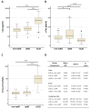
Results: CSF t-tau showed an AUC of 0.942 in the discrimination of sCJD from AD and 0.918 in the discrimination from rpAD. The tau ratio showed significantly higher AUCs (p < 0.001) of 0.992 versus non-rpAD and 0.990 versus rpAD. In the neurochemistry cohort, prion diseases accounted for only 25% of very high CSF t-tau values. High tau-ratios were observed in CJD, but also in non-neurodegenerative diseases.
Conclusions: CSF t-tau is a reliable biomarker for sCJD, but false positive results may occur, especially in rpAD and acute encephalopathies. The t-tau/p-tau ratio may improve the diagnostic accuracy in centers where specific biomarkers are not available.
Keywords: Alzheimer’s disease; Creutzfeldt-Jakob disease; biomarker; cerebrospinal fluid; rapidly-progressive dementia; tau; tau-ratio.
Conflict of interest statement
J.W. has been an honorary speaker for Actelion, Amgen, Beijing Yibai Science and Technology Ltd., Janssen Cilag, Med Update GmbH, Pfizer, Roche Pharma, and has been a member of the advisory boards of Abbott, Biogen, Boehringer Ingelheim, Lilly, MSD Sharp and Dohme, and Roche Pharma and receives fees as a consultant for Immunogenetics and Roboscreen. All authors declare no conflict of interest. The funders had no role in the design of the study; in the collection, analyses, or interpretation of data; in the writing of the manuscript, or in the decision to publish the results.
Figures


Similar articles
-
Stratification by Genetic and Demographic Characteristics Improves Diagnostic Accuracy of Cerebrospinal Fluid Biomarkers in Rapidly Progressive Dementia.J Alzheimers Dis. 2016 Oct 18;54(4):1385-1393. doi: 10.3233/JAD-160267. J Alzheimers Dis. 2016. PMID: 27589519
-
Clinical value of novel blood-based tau biomarkers in Creutzfeldt-Jakob disease.Alzheimers Dement. 2025 Feb;21(2):e14422. doi: 10.1002/alz.14422. Epub 2024 Dec 6. Alzheimers Dement. 2025. PMID: 39641397 Free PMC article.
-
Association of cerebrospinal fluid prion protein levels and the distinction between Alzheimer disease and Creutzfeldt-Jakob disease.JAMA Neurol. 2015 Mar;72(3):267-75. doi: 10.1001/jamaneurol.2014.4068. JAMA Neurol. 2015. PMID: 25559883
-
Rapidly Progressive Alzheimer's Disease: Contributions to Clinical-Pathological Definition and Diagnosis.J Alzheimers Dis. 2018;63(3):887-897. doi: 10.3233/JAD-171181. J Alzheimers Dis. 2018. PMID: 29710713 Review.
-
Cerebrospinal fluid brain-derived proteins in the diagnosis of Alzheimer's disease and Creutzfeldt-Jakob disease.Neuropathol Appl Neurobiol. 2002 Dec;28(6):427-40. doi: 10.1046/j.1365-2990.2002.t01-2-00427.x. Neuropathol Appl Neurobiol. 2002. PMID: 12445159 Review.
Cited by
-
CSF Biomarkers in the Early Diagnosis of Mild Cognitive Impairment and Alzheimer's Disease.Int J Mol Sci. 2023 May 19;24(10):8976. doi: 10.3390/ijms24108976. Int J Mol Sci. 2023. PMID: 37240322 Free PMC article. Review.
-
The Importance of Long-term Partner Observation in Cognitive Evaluation: A Very Early Creutzfeldt-Jakob Disease in a Patient with Mild Cognitive Impairment.Curr Alzheimer Res. 2024;21(3):214-218. doi: 10.2174/0115672050309694240708052535. Curr Alzheimer Res. 2024. PMID: 39041276
-
Tau and neurofilament light-chain as fluid biomarkers in spinocerebellar ataxia type 3.Eur J Neurol. 2022 Aug;29(8):2439-2452. doi: 10.1111/ene.15373. Epub 2022 May 26. Eur J Neurol. 2022. PMID: 35478426 Free PMC article.
-
An Atypical Case of Creutzfeldt-Jakob Syndrome Presenting with Cacosmia and Amyloid Positivity.J Alzheimers Dis Rep. 2024 Jul 23;8(1):1105-1110. doi: 10.3233/ADR-230173. eCollection 2024. J Alzheimers Dis Rep. 2024. PMID: 39434818 Free PMC article.
-
Diagnostic Utility of Cerebrospinal Fluid Biomarkers in Patients with Rapidly Progressive Dementia.Ann Neurol. 2024 Feb;95(2):299-313. doi: 10.1002/ana.26822. Epub 2023 Nov 10. Ann Neurol. 2024. PMID: 37897306 Free PMC article.
References
-
- Ladogana A., Puopolo M., Croes E.A., Budka H., Jarius C., Collins S., Klug G.M., Sutcliffe T., Giulivi A., Alperovitch A., et al. Mortality from Creutzfeldt-Jakob disease and related disorders in Europe, Australia, and Canada. Neurology. 2005;64:1586–1591. doi: 10.1212/01.WNL.0000160117.56690.B2. - DOI - PubMed
-
- Parchi P., Giese A., Capellari S., Brown P., Schulz-Schaeffer W., Windl O., Zerr I., Budka H., Kopp N., Piccardo P., et al. Classification of sporadic Creutzfeldt-Jakob disease based on molecular and phenotypic analysis of 300 subjects. Ann. Neurol. 1999;46:224–233. doi: 10.1002/1531-8249(199908)46:2<224::AID-ANA12>3.0.CO;2-W. - DOI - PubMed
-
- WHO . Global Surveillance, Diagnosis, and Therapy of Human Transmissible Spongiform Encephalopathies: Report of WHO Consultation. WHO; Geneva, Switzerland: 1998.
Publication types
MeSH terms
Substances
Supplementary concepts
LinkOut - more resources
Full Text Sources
Medical

