. 2022 Feb 2;14(2):310.
doi: 10.3390/v14020310.

Salmon Erythrocytes Sequester Active Virus Particles in Infectious Salmon Anaemia

Affiliations

Salmon Erythrocytes Sequester Active Virus Particles in Infectious Salmon Anaemia

Johanna Hol Fosse et al. Viruses. .

Abstract

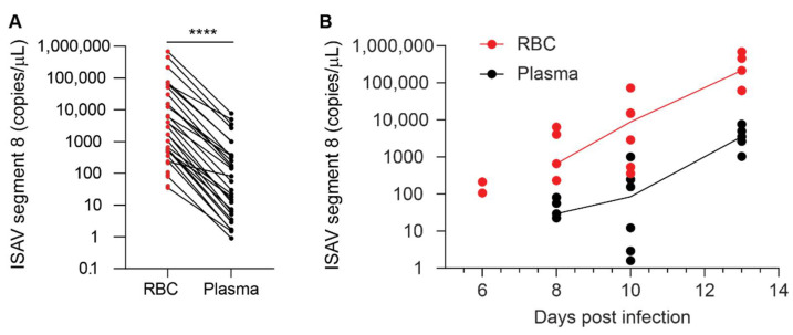
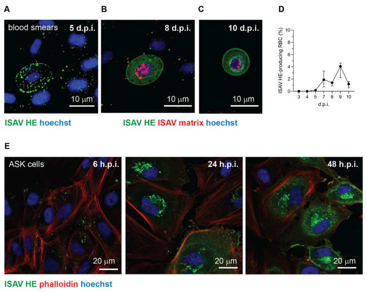
Infectious salmon anaemia virus (ISAV) binds circulating Atlantic salmon erythrocytes, but the relevance of this interaction for the course of infection and development of disease remains unclear. We here characterise ISAV-erythrocyte interactions in experimentally infected Atlantic salmon and show that ISAV-binding to erythrocytes is common and precedes the development of disease. Viral RNA and infective particles were enriched in the cellular fraction of blood. While erythrocyte-associated ISAV remained infectious, erythrocytes dose-dependently limited the infection of cultured cells. Surprisingly, immunostaining of blood smears revealed expression of ISAV proteins in a small fraction of erythrocytes in one of the examined trials, confirming that ISAV can be internalised in this cell type and engage the cellular machinery in transcription and translation. However, viral protein expression in erythrocytes was rare and not required for development of disease and mortality. Furthermore, active transcription of ISAV mRNA was higher in tissues than in blood, supporting the assumption that ISAV replication predominantly takes place in endothelial cells. In conclusion, Atlantic salmon erythrocytes bind ISAV and sequester infective virus particles during infection, but do not appear to significantly contribute to ISAV replication. We discuss the implications of our findings for infection dynamics and pathogenesis of infectious salmon anaemia.

Keywords: adsorption; decoy; isavirus; nucleated erythrocyte; orthomyxovirus; red blood cell; viral replication.


Conflict of interest statement

The authors declare no conflict of interest. The funders had no role in the design of the study; in the collection, analyses, or interpretation of data; in the writing of the manuscript, or in the decision to publish the results.

Figures

Figure 1
Figure 1
ISAV binds Atlantic salmon erythrocyte membrane glycoproteins. Plasma membrane-enriched erythrocyte lysates were separated by gel electrophoresis under denaturing conditions and blotted to nitrocellulose membranes. (A) Periodic acid-schiff staining to visualise glycoproteins. The numbering to the left indicates the weight of the molecular marker in the left lane. (B,C) Virus binding assay to blotted membranes from parallel runs, visualising bound antigen by detection of ISAV HE. (B) Arrow points to the predominant ISAV-binding glycoprotein-containing band; star indicates a second ISAV-binding glycoprotein-containing band; in contrast, two ISAV-binding bands at 35 and 45 kDa indicated by open arrowheads, did not have obvious corresponding PAS bands. (C) Prior saponification of the blotted membrane obliterated all ISAV binding. (D) Total protein staining of representative gel. Images show full-length lanes representative of three independent experiments. Original images have been uploaded as supporting information.
Figure 2
Figure 2
Cumulative mortality in the two ISAV infection trials used in this study. Materials analysed in this study were harvested from two independent ISAV infection trials conducted at different locations and in different years. (A) Cumulative mortality in the 2018 trial (results have been published previously [24], but are included for reference). (B) Cumulative mortality in the 2020 trial.
Figure 3
Figure 3
ISAV coats erythrocytes in experimentally infected Atlantic salmon. (A,B) A reduction in haematocrit was observed in both the 2018 (A) and 2020 (B) trials. Dots represent individual fish, solid lines connect median values, and dotted lines indicate the range of values in the non-infected fish group. (C) ISAV segment 8 qPCR in blood sampled at the peak of viraemia (12 + 13 d.p.i and 13 d.p.i., respectively) in the 2018 and 2020 trials. Dots represent individual fish, bars show median values. ** p = 0.0095, Mann–Whitney U. (D) Viral infection dynamics in full blood in the 2018 and 2020 trials. Dots represent individual fish, lines connect median values. (E) Wide-field micrograph of typical appearance of immunostaining for ISAV HE (green) in blood smear from infected fish (here: 5 d.p.i., 2018 trial). Nuclei are shown in blue. Image signal was enhanced in ImageJ [29] (version 2.1.0/1.53c; Java 1.8.0_172 [64-bit]) using the multiply function (process > maths > multiply). (F) The percentage of HE-positive erythrocytes at the peak of viraemia in both trials. Bars show median +/− 95% confidence intervals of manual counts from 10 microscope fields. **** p < 0.0001, Mann–Whitney U. (G,H) Osmotic fragility of erythrocytes in the infection trials. The graphs show mean erythrocyte lysis at each NaCl concentration +/− SEM of 3 (2018) or 5 (2020) individual infected fish harvested at 9 d.p.i (2018) or 13 d.p.i. (2020), compared to 8 (2018) or 5 (2020) non-infected controls. p-values give the significance of the difference between control and infected fish, as assessed by a RM 2-way ANOVA with the Geisser Greenhouse correction.
Figure 4
Figure 4
ISAV is enriched in the cellular fraction of blood. qPCR of density gradient-purified erythrocytes (RBC, shown in red) and plasma (black) from individual fish in the 2020 trial showing (A) the relationship between ISAV mRNA in RBC and plasma in all individual fish and (B) the relationship of ISAV mRNA in RBC and plasma over the time course of the experimental infection. Dots represent individual fish. (A) Lines connect values from the same fish. **** p < 0.0001, Wilcoxon matched-pairs signed rank test. (B) Lines connect median values within each group. The values on the y-axis have been adjusted for the difference in the sampled volume of RBC and plasma.
Figure 5
Figure 5
Occasionally, a subset of erythrocytes in infected Atlantic salmon express ISAV proteins. (AC) Confocal micrographs of immunostained blood smears from ISAV-infected fish in the 2018 trial. (A) Example of the most frequently observed pattern with extensive coating of erythrocytes with HE-positive particles. (B,C) Examples of the few erythrocytes (B) and rare rounded erythrocyte-like cells (C) that express ISAV HE and matrix proteins. Images show maximum intensity projections from z-stacks. (D) Percentage of HE-expressing erythrocytes in the 2018 trial. The graph shows medians (dots) and 95% confidence intervals (bars) calculated from manual counts of 10 microscope fields, 3 fish per time point. (E) Confocal micrographs of immunostained ISAV-infected ASK cells illustrate the typical change in signal pattern as the infectious cycle progresses. In the initial stage of infection, staining reveals a bright punctuate pattern (left panel). Once cells start to express ISAV HE protein, a bright perinuclear signal appears in the region of the Golgi apparatus (middle panel). Finally, extensive cytoplasmic and membrane staining is present in the late stage of infection (right panel). Images in panel E were obtained in a single z plane.
Figure 6
Figure 6
ISAV predominantly replicates in endothelial cells of solid organs. (A,B) Active viral transcription was estimated by calculating the ratio between ISAV segment 7 mRNA and RNA. Dots show values in individual fish, bars show median and interquartile ranges. (A) ISAV transcription in heart and kidney was higher than in RBC, plasma, and purified ISAV (13 d.p.i., 2020 trial). Stars indicate the difference between active transcription in RBC compared to heart (** p = 0.0087) and head kidney (** p = 0.0079), Mann–Whitney U. (B) ISAV transcription in full blood from fish harvested 12 and 13 d.p.i. in the 2018 trial (2018) and in erythrocytes harvested 13 d.p.i. in the 2020 trial (2020), detecting no significant difference between trials. (C,D) Micrographs showing immunostaining for ISAV nucleoprotein (NP, magenta) in formalin-fixed paraffin-embedded heart (C) and head kidney (D) from fish harvested 13 d.p.i. in the 2020 trial. Arrows point to ISAV NP-positive endothelial cells. Scalebars are 100 µm. (EG) RBC from healthy fish were infected ex vivo with ISAV and harvested 1, 24, 48, and 72 h post infection (h.p.i.). qPCR was used to measure (E) ISAV segment 8, (F) IFNa, and (G) Mx transcripts. ELF-1α was used as reference for calculating fold change in F and G by the ΔΔCT method. The graphs show means +/− standard deviations from RBC from 4 individual fish. A representative time curve of results from infected ASK cells is included as reference.
Figure 7
Figure 7
Erythrocytes sequester infective ISAV particles and inhibit serial infection of cultured cells. (A) Density gradient-purified erythrocytes (RBC) and plasma samples were harvested from ISAV-infected fish (13 d.p.i. in the 2020 trial) and inoculated on ASK cells. Data points represent samples from individual fish. ** p = 0.079, Mann–Whitney U. (B) Serially diluted RBC from healthy fish were added to infected ASK cells 24 h p.i. and incubated another 24 h before supernatants were transferred to new ASK cells. Infection of ASK cells 72 h.p.i. was measured by immunostaining for ISAV NP and automated quantification of the number of infected cells (Spectramax i3x plate reader, minimax 300 Imaging Cytometer module, Molecular devices, CA, USA). The infectivity graphs show medians (dots) and 95% confidence intervals (bars), representative of two independent experiments. *** p < 0.001, ** p < 0.01, one sample t test. (C) Serially diluted RBC from healthy fish were incubated with ISAV. The total levels of ISAV RNA associated with the diminishing RBC pellets were measured by qPCR (red, left y-axis). ISAV RNA relative to the RBC content in the pellet was calculated by the ∆∆CT method, using ELF1α as reference gene and the highest dilution of RBC with ELF1α CT < 35 (considered the reliable limit of detection) as reference sample (black, right y-axis, open circle indicates ELF1α CT > 35). Data show the means of technical duplicates representative of two independent experiments.

Similar articles

Cited by

References

    1. Aamelfot M., Christiansen D.H., Dale O.B., McBeath A., Benestad S.L., Falk K. Localised Infection of Atlantic Salmon Epithelial Cells by HPR0 Infectious Salmon Anaemia Virus. PLoS ONE. 2016;11:e0151723. doi: 10.1371/journal.pone.0151723. - DOI - PMC - PubMed
    1. Christiansen D.H., McBeath A.J.A., Aamelfot M., Matejusova I., Fourrier M., White P., Petersen P.E., Falk K. First field evidence of the evolution from a non-virulent HPR0 to a virulent HPR-deleted infectious salmon anaemia virus. J. Gen. Virol. 2017;98:595–606. doi: 10.1099/jgv.0.000741. - DOI - PubMed
    1. Aamelfot M., Dale O.B., Falk K. Infectious salmon anaemia-pathogenesis and tropism. J. Fish Dis. 2014;37:291–307. doi: 10.1111/jfd.12225. - DOI - PubMed
    1. Aquatic Animal Health Code. [(accessed on 31 August 2021)]. Available online: https://www.oie.int/en/what-we-do/standards/codes-and-manuals/aquatic-co...
    1. Evensen O., Thorud K.E., Olsen Y.A. A morphological study of the gross and light microscopic lesions of infectious anaemia in Atlantic salmon (Salmo salar) Res. Vet. Sci. 1991;51:215–222. doi: 10.1016/0034-5288(91)90017-I. - DOI - PubMed

Publication types

MeSH terms

LinkOut - more resources