Review
. 2022 Feb 5;14(2):324.
doi: 10.3390/v14020324.

Visualization of Retroviral Gag-Genomic RNA Cellular Interactions Leading to Genome Encapsidation and Viral Assembly: An Overview

Affiliations
Review

Visualization of Retroviral Gag-Genomic RNA Cellular Interactions Leading to Genome Encapsidation and Viral Assembly: An Overview

Serena Bernacchi. Viruses. .

Abstract

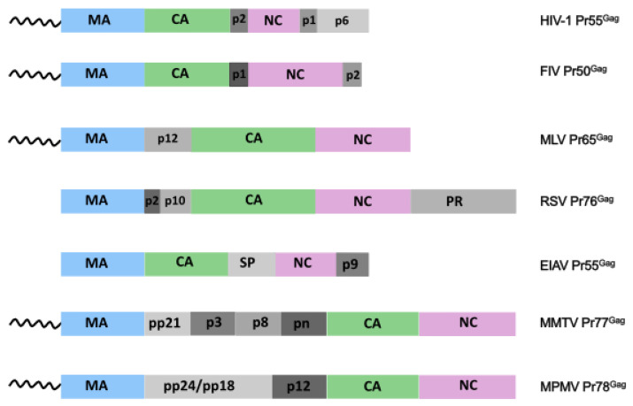
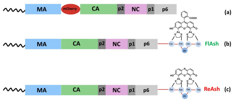
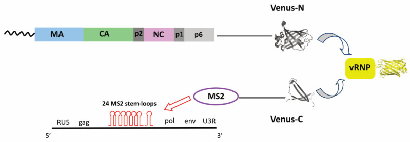
Retroviruses must selectively recognize their unspliced RNA genome (gRNA) among abundant cellular and spliced viral RNAs to assemble into newly formed viral particles. Retroviral gRNA packaging is governed by Gag precursors that also orchestrate all the aspects of viral assembly. Retroviral life cycles, and especially the HIV-1 one, have been previously extensively analyzed by several methods, most of them based on molecular biology and biochemistry approaches. Despite these efforts, the spatio-temporal mechanisms leading to gRNA packaging and viral assembly are only partially understood. Nevertheless, in these last decades, progress in novel bioimaging microscopic approaches (as FFS, FRAP, TIRF, and wide-field microscopy) have allowed for the tracking of retroviral Gag and gRNA in living cells, thus providing important insights at high spatial and temporal resolution of the events regulating the late phases of the retroviral life cycle. Here, the implementation of these recent bioimaging tools based on highly performing strategies to label fluorescent macromolecules is described. This report also summarizes recent gains in the current understanding of the mechanisms employed by retroviral Gag polyproteins to regulate molecular mechanisms enabling gRNA packaging and the formation of retroviral particles, highlighting variations and similarities among the different retroviruses.

Keywords: Gag precursor; cellular trafficking; fluorescent labelling; genomic RNA packaging; live-cells microscopy; plasma membrane; retroviral assembly; retrovirus; ribonucleoprotein complex; specific interactions.


Conflict of interest statement

No conflict of interest declared.

Figures

Figure 1
Figure 1
Retroviral Gag precursors. Three domains are found in all the retroviral Gag proteins: matrix (MA, light blue) at the N-terminus, capsid (CA, green) and nucleocapsid (NC, pink). In addition to those main domains, retroviral Gag also possess other small domains (various shades of grey) located at various positions which display different activities.
Figure 2
Figure 2
Different labelling strategies allow the detection of Gag precursors in cells. (a) Gag fusion to FPs such as m-Cherry was, for example, used to analyze the trafficking of HIV-1 Gag-gRNA complexes to the PM where the assembly occurs [21]. Membrane-permeable biarsenical compounds named (b) FlAsH, whose structure is based around a fluorescein core, and (c) ReAsH, which is a derivative of resorufin, were also used to observe HIV-1 Gag dynamics [20]. In this case, Gag labelling was achieved by adding a linear tetraCys motif (TC-tag), where XX is usually Pro-Gly [74]. Importantly, this labelling is highly specific, as Gag becomes fluorescent only when the biarsenical compounds bind the target.
Figure 3
Figure 3
In BiFC assays, the two halves of Venus fluorescent protein are fused to two interacting proteins of interest, as for example Gag and MS2 proteins [147]. One of the most widely used strategies to monitor gRNA trafficking in cells consists in the insertion of 24 stem-loop sequences in the target RNA which are then recognized with high affinity and specificity by the bacteriophage MS2 coat protein. While the two halves of Venus protein are non-fluorescent, the interaction of the partners tethers the fused fluorescent fragments in proximity, which facilitates the restoration of Venus fluorescence. BiFC is a reliable and sensitive approach that also revealed molecular interactions in other viral contexts, such as the Herpes virus [148].

Similar articles

Cited by

References

    1. Mailler E., Bernacchi S., Marquet R., Paillart J.-C., Vivet-Boudou V., Smyth R. The Life-Cycle of the HIV-1 Gag–RNA Complex. Viruses. 2016;8:248. doi: 10.3390/v8090248. - DOI - PMC - PubMed
    1. Rein A., Datta S.A.K., Jones C.P., Musier-Forsyth K. Diverse Interactions of Retroviral Gag Proteins with RNAs. Trends Biochem. Sci. 2011:S0968000411000545. doi: 10.1016/j.tibs.2011.04.001. - DOI - PMC - PubMed
    1. Olson E.D., Musier-Forsyth K. Retroviral Gag Protein-RNA Interactions: Implications for Specific Genomic RNA Packaging and Virion Assembly. Semin. Cell Dev. Biol. 2019;86:129–139. doi: 10.1016/j.semcdb.2018.03.015. - DOI - PMC - PubMed
    1. Dubois N., Marquet R., Paillart J.-C., Bernacchi S. Retroviral RNA Dimerization: From Structure to Functions. Front. Microbiol. 2018;9:527. doi: 10.3389/fmicb.2018.00527. - DOI - PMC - PubMed
    1. Lever A.M.L. Advances in Pharmacology. Volume 48. Elsevier; Amsterdam, The Netherlands: 2000. HIV RNA Packaging and Lentivirus-Based Vectors; pp. 1–28. - PubMed

Publication types

Substances

LinkOut - more resources