. 2022 Feb 8;14(2):349.
doi: 10.3390/v14020349.

Isolation, Characterization, and Molecular Detection of Porcine Sapelovirus

Affiliations

Isolation, Characterization, and Molecular Detection of Porcine Sapelovirus

Yassein M Ibrahim et al. Viruses. .

Abstract

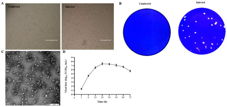
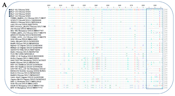
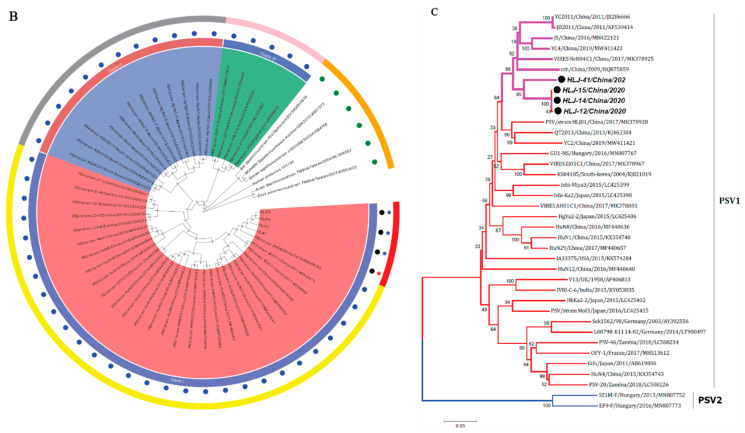
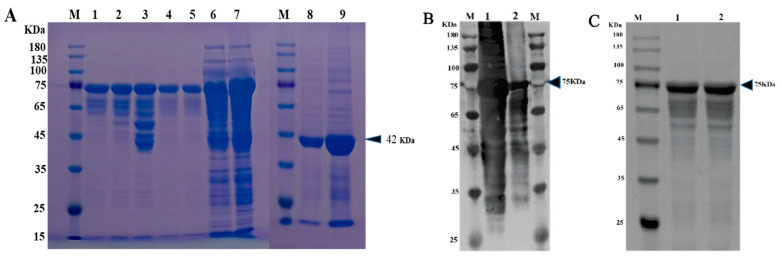
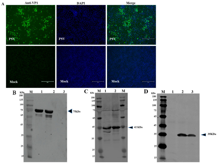
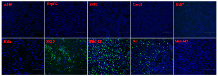
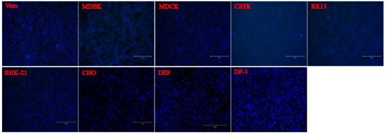
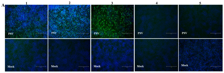
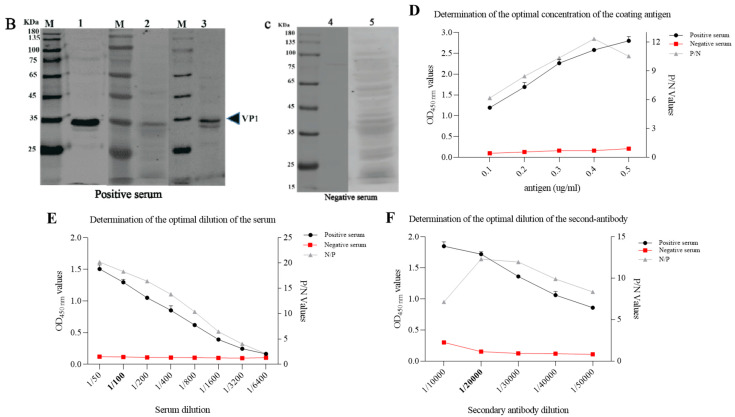
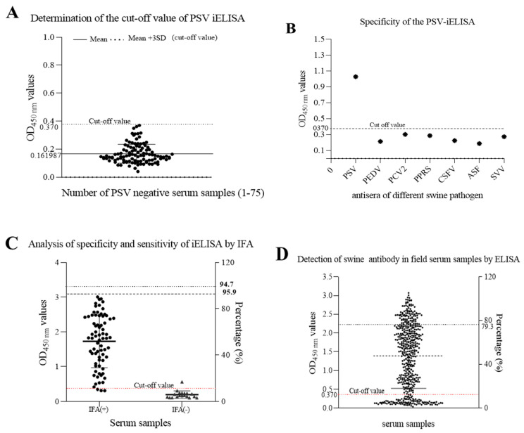
Porcine sapelovirus (PSV) is an important emerging pathogen associated with a wide variety of diseases in swine, including acute diarrhoea, respiratory distress, skin lesions, severe neurological disorders, and reproductive failure. Although PSV is widespread, serological assays for field-based epidemiological studies are not yet available. Here, four PSV strains were recovered from diarrheic piglets, and electron microscopy revealed virus particles with a diameter of ~32 nm. Analysis of the entire genome sequence revealed that the genomes of PSV isolates ranged 7569-7572 nucleotides in length. Phylogenetic analysis showed that the isolated viruses were classified together with strains from China. Additionally, monoclonal antibodies for the recombinant PSV-VP1 protein were developed to specifically detect PSV infection in cells, and we demonstrated that isolated PSVs could only replicate in cells of porcine origin. Using recombinant PSV-VP1 protein as the coating antigen, we developed an indirect ELISA for the first time for the detection of PSV antibodies in serum. A total of 516 swine serum samples were tested, and PSV positive rate was 79.3%. The virus isolates, monoclonal antibodies and indirect ELISA developed would be useful for further understanding the pathophysiology of PSV, developing new diagnostic assays, and investigating the epidemiology of the PSV.

Keywords: ELISA; characterization; isolation; monoclonal antibodies; porcine sapelovirus; prevalence.


Conflict of interest statement

The authors declare no conflict of interest.

Figures

Figure 1
Figure 1
Isolation and identification of PSV. (A) Cytopathic effects in PSV-infected PK15 cells at 24 hpi. (B) Production of plaques of the PSV isolate in PK15 cells. (C) Picornavirus-like particles under TEM. (D) Growth kinetic of PSV.
Figure 2
Figure 2
Sequence analysis of the isolated PSV strains. (A) Alignment of 3′-partial amino acid sequences of VP1 of PSV strains. Sequences in boxes showed the hypervariable region in the C-terminus of VP1. The strains identified in the present study were indicated by black dots; (-), missing amino acids. (B,C) Phylogenetic analysis of PSV based on the complete nucleotide sequence and VP1 gene respectively. The trees were generated by using MEGA v.6.0 with the neighbor-joining method with the Kimura 2-parameter with 1000 bootstrap replication. The black circle indicates PSV isolated in this study. The scale bars represent the number of substitutions per site.
Figure 2
Figure 2
Sequence analysis of the isolated PSV strains. (A) Alignment of 3′-partial amino acid sequences of VP1 of PSV strains. Sequences in boxes showed the hypervariable region in the C-terminus of VP1. The strains identified in the present study were indicated by black dots; (-), missing amino acids. (B,C) Phylogenetic analysis of PSV based on the complete nucleotide sequence and VP1 gene respectively. The trees were generated by using MEGA v.6.0 with the neighbor-joining method with the Kimura 2-parameter with 1000 bootstrap replication. The black circle indicates PSV isolated in this study. The scale bars represent the number of substitutions per site.
Figure 3
Figure 3
Expression and purification of PSV-VP1 protein in E. coli. (A) SDS-PAGE analysis of the MBP-tagged recombinant VP1-protein expression (~75 KDa). Lane M, protein marker; Lanes 1–5, purified bacterial cell lysates of MBP-tagged VP1 at different concentrations; Lines 6 and 7, unpurified bacterial cell lysate of MBP-tagged VP1; Line 8, sediment of bacterial cell lysate of empty vector, Line 9, supernatant of bacterial cell lysate of =empty vector (~42 KDa). (B) Western blot analysis of unpurified recombinant VP1-protein using anti-MBP tag antibody. Lane 1, 0.2 mg/mL of unpurified VP1 protein; Lane 2, 0.1 mg/mL of unpurified VP1 protein. (C) Western blot analysis of purified VP1-recombinant protein using anti-MBP tag antibody. Lane 1, 0.1 mg/mL of purified VP1 protein; Lane 2, 0.2 mg/mL of purified VP1 protein.
Figure 4
Figure 4
Identification of mAb against VP1 of PSV. (A) Identification of mAb against PSV-VP1 by IFA. PK15 cells infected with PSV at MOI of 0.01. IFA was performed using generated mAb against VP1. (B) Western blot analysis of the purified recombinant protein from E. Coli. Lane M, protein marker; Lane 1, 0.1 mg/mL purified MBP-tagged VP1-recombinant protein; Lane 2, 0.2 mg/mL MBP-tagged VP1-recombinant protein; Lane 3, purified MBP. (C) Western blot analysis of the expressed MBP in empty vector. Lane M, protein marker; Lane 1, sediment of bacterial cell lysate, Line 2, supernatant of bacterial cell lysate. (D) Western blot analysis of PK15 cells infected with PSV. Lane M, protein marker; Lane 1, mock cells; Lane 2, PK15 cells infected with PSV; Lane 3, ST cells infected with PSV.
Figure 5
Figure 5
Susceptibility of PSV to cell lines derived from various species. Cell lines of Human (A549, HeLa, Huh-7, HepG2, Coca2 and 293T), Swine (PK15, IPEC-J2, and ST), Monkeys (Marc-145 and Vero), Bovine (MDBK), Canine (MDCK), Feline (CRFK), Rabbit (RK13), Hamster (BHK-21 and CHO), Duck (DEF) and Chicken (DF-1) were infected with PSV at an MOI of 0.01, and fixed at 24 hpi. Cell monolayers were determined by IFA using PSV mAb.
Figure 5
Figure 5
Susceptibility of PSV to cell lines derived from various species. Cell lines of Human (A549, HeLa, Huh-7, HepG2, Coca2 and 293T), Swine (PK15, IPEC-J2, and ST), Monkeys (Marc-145 and Vero), Bovine (MDBK), Canine (MDCK), Feline (CRFK), Rabbit (RK13), Hamster (BHK-21 and CHO), Duck (DEF) and Chicken (DF-1) were infected with PSV at an MOI of 0.01, and fixed at 24 hpi. Cell monolayers were determined by IFA using PSV mAb.
Figure 6
Figure 6
Optimization of reaction conditions of iELISA. (A) Verification of PSV-negative and positive swine sera by IFA. PK15 cells were inoculated with PSV at MOI of 0.01. IFA was performed using swine positive serum # 1, 2, and 3, and negative serum # 4 and 5. These are the representative sera of all the tested sera. (B,C) Confirmation of PSV positive and negative serum by Western blot. Protein lysates of PK15 cells infected with PSV were detected with PSV positive and negative swine serum respectively. (DF) Optimization of the concentrations of coating antigen, serum dilutions and second antibody respectively.
Figure 6
Figure 6
Optimization of reaction conditions of iELISA. (A) Verification of PSV-negative and positive swine sera by IFA. PK15 cells were inoculated with PSV at MOI of 0.01. IFA was performed using swine positive serum # 1, 2, and 3, and negative serum # 4 and 5. These are the representative sera of all the tested sera. (B,C) Confirmation of PSV positive and negative serum by Western blot. Protein lysates of PK15 cells infected with PSV were detected with PSV positive and negative swine serum respectively. (DF) Optimization of the concentrations of coating antigen, serum dilutions and second antibody respectively.
Figure 7
Figure 7
Detection of anti-PSV antibodies in swine serum by the developed iELISA. (A) seventy-five PSV-negative serum samples were tested using iELISA and the mean OD450 value of PSV-negative serum plus three standard deviations (SDs) were used to calculate the cutoff value. (B) Identification of iELISA specificity by cross-reaction test. Positive swine serum against PSV, PEDV, PCV2, PRRS, CSFV, ASFV and SVV were tested by iELISA. The average of OD450 values were calculated to determine the tested serum according to the cut-off value. (C) The sensitivity and specificity of the developed ELISA were assessed based on the IFA result. (D) Detection of anti-PSV antibodies from field swine serum samples by the developed iELISA.

Similar articles

Cited by

References

    1. Li Y., Du L., Jin T., Cheng Y., Zhang X., Jiao S., Huang T., Zhang Y., Yan Y., Gu J. Characterization and epidemiological survey of porcine sapelovirus in China. Vet. Microbiol. 2019;232:13–21. doi: 10.1016/j.vetmic.2019.02.017. - DOI - PubMed
    1. Yang T., Yu X., Yan M., Luo B., Li R., Qu T., Luo Z., Ge M., Zhao D. Molecular characterization of Porcine sapelovirus in Hunan, China. J. Gen. Virol. 2017;98:2738–2747. doi: 10.1099/jgv.0.000951. - DOI - PubMed
    1. Oberste M.S., Maher K., Kilpatrick D.R., Pallansch M.A. Molecular evolution of the human enteroviruses: Correlation of serotype with VP1 sequence and application to picornavirus classification. J. Virol. 1999;73:1941–1948. doi: 10.1128/JVI.73.3.1941-1948.1999. - DOI - PMC - PubMed
    1. Son K.-Y., Kim D.-S., Kwon J., Choi J.-S., Kang M.-I., Belsham G.J., Cho K.-O. Full-length genomic analysis of Korean porcine Sapelovirus strains. PLoS ONE. 2014;9:e107860. doi: 10.1371/journal.pone.0107860. - DOI - PMC - PubMed
    1. Mateu M.G. Antibody recognition of picornaviruses and escape from neutralization: A structural view. Virus Res. 1995;38:1–24. doi: 10.1016/0168-1702(95)00048-U. - DOI - PubMed

Publication types

MeSH terms

Substances

LinkOut - more resources