Review
. 2022 Feb 8;14(2):351.
doi: 10.3390/v14020351.

Known Cellular and Receptor Interactions of Animal and Human Coronaviruses: A Review

Affiliations
Review

Known Cellular and Receptor Interactions of Animal and Human Coronaviruses: A Review

Holly Everest et al. Viruses. .

Abstract

This article aims to review all currently known interactions between animal and human coronaviruses and their cellular receptors. Over the past 20 years, three novel coronaviruses have emerged that have caused severe disease in humans, including SARS-CoV-2 (severe acute respiratory syndrome virus 2); therefore, a deeper understanding of coronavirus host-cell interactions is essential. Receptor-binding is the first stage in coronavirus entry prior to replication and can be altered by minor changes within the spike protein-the coronavirus surface glycoprotein responsible for the recognition of cell-surface receptors. The recognition of receptors by coronaviruses is also a major determinant in infection, tropism, and pathogenesis and acts as a key target for host-immune surveillance and other potential intervention strategies. We aim to highlight the need for a continued in-depth understanding of this subject area following on from the SARS-CoV-2 pandemic, with the possibility for more zoonotic transmission events. We also acknowledge the need for more targeted research towards glycan-coronavirus interactions as zoonotic spillover events from animals to humans, following an alteration in glycan-binding capability, have been well-documented for other viruses such as Influenza A.

Keywords: SARS-CoV-2; cleavage; coronavirus; glycan; haemagglutinin-esterase; host interaction; omicron; receptor-binding; sialic acid; spike protein.


Conflict of interest statement

The authors declare no conflict of interest.

Figures

Figure 1
Figure 1
A midpoint rooted circular cladogram of representative coronavirus spike glycoprotein sequences across the alpha-, beta-, gamma- and delta-genera. Representative strains from Genbank were used (Accession Numbers outline in Table 1). MUSCLE [22] was used to align the sequences in MEGA X [23]. A Neighbour-Joining cladogram was generated, which highlights the specific coronavirus genera and is coloured accordingly. The genera are also annotated in bold font denoted by Greek symbol (alpha-α, beta-β, delta-δ, gamma-γ).
Figure 5
Figure 5
A midpoint rooted cladogram of representative coronavirus S glycoprotein sequences across the alpha-, beta-, gamma- and delta-genres. Representative strains from Genbank were used (Accesion Numbers outlined in Table 1). MUSCLE [22] was used to align the sequences in MEGA X [23]. A Neighbour-Joining cladogram was generated and the coronavirus genera denoted by Greek symbol (alpha-α, beta-β, delta-δ, gamma-γ). A linear schematic of the S glycoprotein S1 domain is also indicated for each virus (NTD in pink and CTD in blue—additional binding domain A of FCoV indicated in green). The receptor bound by the relevant domain is annotated accordingly.
Figure 6
Figure 6
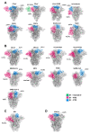
Spike glycoprotein schematic diagrams with putative receptor-binding domains indicated (as per Figure 5, NTD in pink and CTD in blue—additional binding domain A of FCoV indicated in green). (A) = alphacoronavirus, (B) = betacoronavirus, (C) = deltacoronavirus, (D) = gammacoronavirus genres. The Genbank accession numbers for the representative sequences used for modelling in PyMol [61], and the RCSB PDB ID used as a SWISS-MODEL [82] backbone are denoted in Table 1.
Figure 7
Figure 7
Structure and linear schematic of HKU-1 HE glycoprotein structure modelled in PyMol [61] using the Cryo-EM structure of HKU-1 [128] (RCSB PDB: 6Y3Y). (A) A single monomer is highlighted with each region of the HE glycoprotein annotated. (B) Linear schematic diagram of HKU-1 HE ectodomain.
Figure 8
Figure 8
A midpoint rooted cladogram of betacoronavirus Lineage A viruses. Representative strains from Genbank were used (Accession Numbers outlined in Table 2). MUSCLE [22] was used to align the sequences in MEGA X [23]. A Neighbour-Joining cladogram was generated. A linear schematic of the HE glycoprotein lectin and esterase domains is also indicated for each virus (Esterase in orange and Lectin in purple). The receptor bound by the relevant domain is annotated accordingly.
Figure 9
Figure 9
HE glycoprotein schematic diagrams with putative lectin binding domains indicated (as per Figure 8, Lectin domain in purple). The Genbank accession numbers for the representative sequences used for modelling in PyMol [61], and the RCSB PDB ID used as a SWISS-MODEL [82] backbone are denoted in Table 2.
Figure 2
Figure 2
Annotated diagrams of the coronavirus virion—the presence of the HE surface protein differentiates the betacoronavirus lineage A viruses (right) from other coronaviruses (left). The surface proteins are also denoted by colour S glycoprotein (purple) and HE glycoprotein (green). Figure Adapted from “Human Coronavirus Structure”, by BioRender.com (2021). Available online: https://app.biorender.com/biorender-templates (accessed on 2 February 2022).
Figure 3
Figure 3
The cellular entry mechanisms of coronaviruses. Route (A) = internalisation via endocytosis, Route (B) = internalisation via membrane fusion. Figure adapted from the “Coronavirus Replication Cycle” by BioRender.com (2021). Available online: https://app.biorender.com/biorender-templates (accessed on 2 February 2022).
Figure 4
Figure 4
Structure and schematic of IBV S glycoprotein in pre-fusion conformation with 2P stabilisation. (A) IBV trimeric structure modelled in PyMol [61] using the Cryo-EM structure of IBV strain M41 [53] (RCSB PDB: 6CVO). (A) single monomer is highlighted with each region of the S glycoprotein annotated. (B) The M41 IBV structure modelled in PyMol with the substitution of two proline amino acid residues allowing for pre-fusion 2P stabilisation. The proline insertions are indicated. (C) Linear schematic diagram of IBV spike ectodomain. Question mark indicates that the exact amino acid residue location of the FP is unidentified.
Figure 10
Figure 10
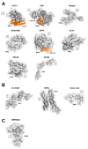
Receptor schematic diagrams with putative binding regions (where known) highlighted in orange, with amino acid residue locations noted. (A) = Protein Receptors, (B) = Sugar Receptors, (C) = Other Binding Factors. The RCSB PDB for the representative models made in PyMol [61] are denoted below the relevant model.

Similar articles

Cited by

References

    1. King A.M., Lefkowitz E., Adams M.J., Carstens E.B. Virus Taxonomy: Ninth Report of the International Committee on Taxonomy of Viruses. Volume 9 Elsevier; Amsterdam, The Netherlands: 2011.
    1. Walker P.J., Siddell S.G., Lefkowitz E.J., Mushegian A.R., Adriaenssens E.M., Dempsey D.M., Dutilh B.E., Harrach B., Harrison R.L., Hendrickson R.C., et al. Changes to virus taxonomy and the Statutes ratified by the International Committee on Taxonomy of Viruses (2020) Arch. Virol. 2020;165:2737–2748. doi: 10.1007/s00705-020-04752-x. - DOI - PubMed
    1. Lefkowitz E.J., Dempsey D.M., Hendrickson R.C., Orton R.J., Siddell S.G., Smith D.B. Virus taxonomy: The database of the International Committee on Taxonomy of Viruses (ICTV) Nucleic Acids Res. 2018;46:D708–D717. doi: 10.1093/nar/gkx932. - DOI - PMC - PubMed
    1. Beaudette F. Cultivation of the virus of infectious bronchitis. J. Am. Vet. Med. Assoc. 1937;90:51–60.
    1. Doyle L.P., Hutchings L.M. A transmissible gastroenteritis in pigs. J. Am. Vet. Med. Assoc. 1946;108:257–259. - PubMed

Publication types

LinkOut - more resources