. 2022 Feb 8;14(2):348.
doi: 10.3390/v14020348.

Structure-Based Discovery of N-Sulfonylpiperidine-3-Carboxamides as Novel Capsid Assembly Modulators for Potent Inhibition of HBV Replication

Affiliations

Structure-Based Discovery of N-Sulfonylpiperidine-3-Carboxamides as Novel Capsid Assembly Modulators for Potent Inhibition of HBV Replication

Yang Yang et al. Viruses. .

Abstract

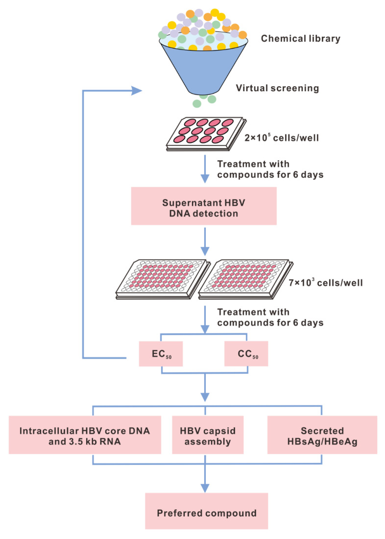
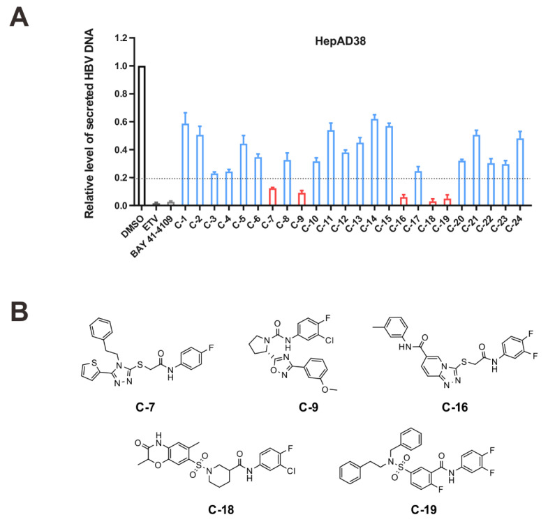
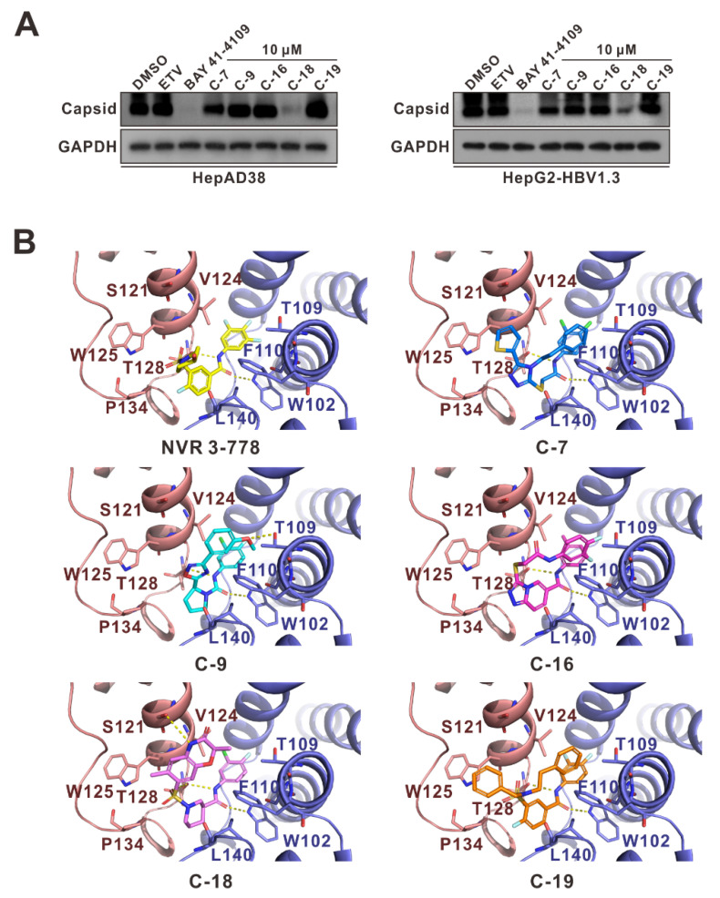
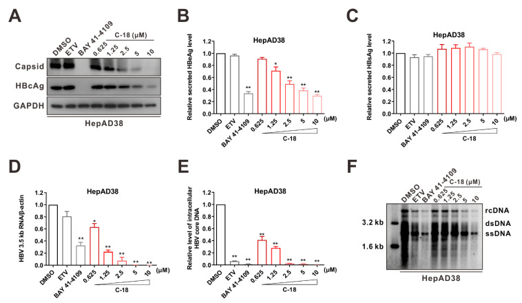
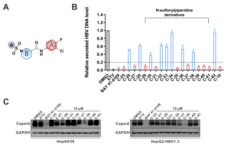
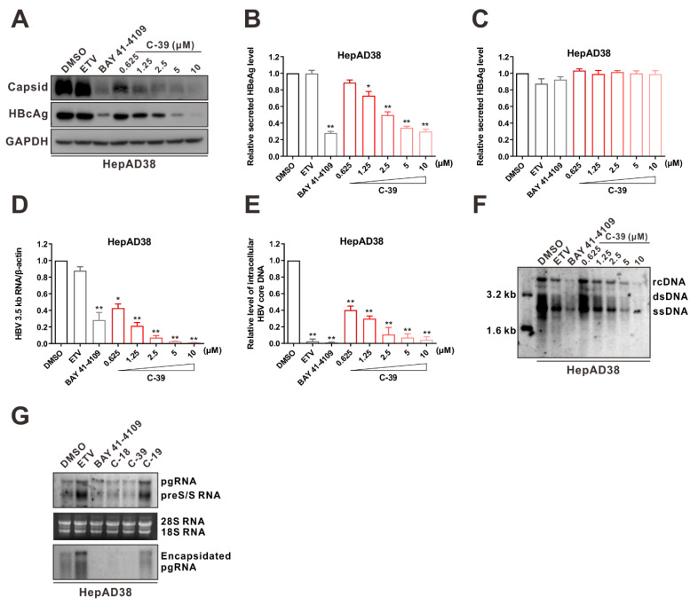
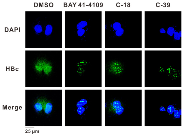
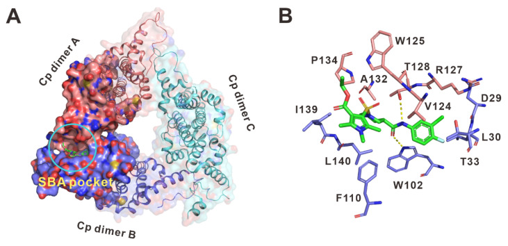
As a key element during HBV replication, a nucleocapsid is considered a promising target for the treatment of chronic hepatitis B. The present study aimed to identify small molecules as novel capsid assembly modulators with antiviral activity. Structure-based virtual screening of an integrated compound library led to the identification of several types of HBV inhibitors. Among these inhibitors, N-sulfonylpiperidine-3-carboxamides (SPCs) potently reduced the amount of secreted HBV DNA. Through structure-activity relationship studies, we identified an SPC derivative, namely, C-39, which exhibited the highest antiviral activity without causing cytotoxicity. Mechanism studies showed that C-39 dose-dependently inhibited the formation of HBV capsid, synthesis of cccDNA, e antigen (HBeAg), viral pregenomic RNA (pgRNA), and HBV DNA levels, thereby restraining HBV replication. In summary, SPCs represent a new class of capsid assembly modulators. Further optimization of SPCs is expected to obtain new antiviral drugs against HBV infection.

Keywords: antiviral activity; assembly modulator; capsid; core protein; hepatitis B virus.


Conflict of interest statement

The authors declare no conflict of interest.

Figures

Figure 1
Figure 1
Schematic depiction of the primary screening workflow. HepAD38 or HepG2-HBV1.3 cells were treated with compounds (20 μM) for 6 days, then viral nucleic acids and proteins were detected.
Figure 2
Figure 2
Screening and identification of novel HBV inhibitors. (A) Effects of the 24 compounds (C-1–C-24, 20 μM) on secreted HBV DNA in HepAD38 cells. Compounds with significant antiviral activity were defined with a threshold of 20% of DMSO control. (B) Structures of the initial hit compounds C-7, C-9, C-16, C-18, and C-19. Data are representative of three independent experiments and are expressed as the mean ± SD.
Figure 3
Figure 3
Effects of initial hits on viral particles and molecular interaction analysis. (A) Effects of five initial hits on intracellular HBV capsid formation. HepAD38 or HepG2-HBV1.3 cells were incubated with C-7, C-9, C-16, C-18, C-19 (10 μM), ETV (25 nM), or BAY 41-4109 (2 μM) for 6 days. HBV core particles were isolated from cell lysates by 1.2% non-denaturing agarose gel electrophoresis and immunoblotting. (B) Putative molecular interactions between HBV core protein dimers and NVR 3-778, C-7, C-9, C-16, C-18, or C-19. Compounds and residues were represented as sticks. Yellow dashed lines indicated the hydrogen bonds.
Figure 4
Figure 4
C-18 inhibits HBV replication in HepAD38 cells. (A) Effects of C-18 on intracellular HBV capsid formation and HBc. HepAD38 cells were incubated with the indicated concentrations of C-18 for 6 days. Capsids and HBV core protein were probed with anti-HBV core antibody, and GAPDH was used as the loading control. (BF) Effects of C-18 on HBV replication. HepAD38 cells were incubated with the indicated concentrations of C-18 for 6 days. Secreted HBeAg (B) and HBsAg (C) were determined using ELISA, intracellular HBV 3.5 kb RNA (D) and HBV DNA (E) were measured using qPCR, and intracellular HBV replication intermediates (RIs) were detected using Southern blot analysis. rcDNA, relaxed circular DNA; dsDNA, double-stranded DNA; ssDNA, single-stranded DNA. (F) ETV (25 nM) and BAY 41-4109 (2 μM) were used as controls. The data in (BE) are representative of three independent experiments and are expressed as mean ± SD (Student’s t-test was used to assess the statistical significance between DMSO group against any other treatment; * p < 0.05, ** p < 0.01).
Figure 5
Figure 5
Effects of the rescreened compounds on HBV replication and capsid formation. (A) The scaffold of C-18. The substituent on the SPC scaffold was represented as an R group. Ring A and ring B are highlighted. (B) Effects of the 18 compounds (C-25–C-42) and C-18 on secreted HBV DNA in HepAD38 cells. The threshold for hit selection was set stringently to a 20% residual level relative to the control (DMSO). Data are representative of three independent experiments and are expressed as the mean ± SD. (C) Effects of nine selected compounds on intracellular HBV capsid formation. HepAD38 and HepG2-HBV1.3 were incubated with nine selected compounds (10 μM), ETV (25 nM), and BAY 41-4109 (2 μM) for 6 days.
Figure 6
Figure 6
C-39 significantly inhibits HBV replication. HepAD38 cells were incubated with the indicated concentrations of C-39 for 6 days. (A) Effects of C-39 on intracellular HBV capsid formation. HBV capsids and core protein were assessed using the anti-HBV core antibody. (BF) Effects of C-39 on HBV replication. Secreted HBeAg (B) and HBsAg (C) were determined using ELISA, intracellular HBV 3.5 kb RNA (D) and core DNA (E) were measured using qPCR, and intracellular HBV replication intermediates (RIs) were assessed using Southern blotting (F). (G) Effects of C-18, C-39, and C-19 (5 μM) on the HBV total RNAs and encapsidated pgRNA were determined using Northern blotting. ETV (25 nM) and BAY 41-4109 (2 μM) were used as controls. The data in (BE) are representative of three independent experiments and are expressed as mean ± SD (Student’s t-test was used to assess the statistical significance between DMSO group against any other treatment; * p < 0.05, ** p < 0.01).
Figure 7
Figure 7
Effects of C-39 on the HBc aggregation. Confocal immunofluorescence microscopy images showing HBV core protein (in green) distribution in HepAD38 cells after 3 days of incubation of DMSO, BAY 41-4109 (2 μM), C-18 (5 μM), and C-39 (5 μM). Nuclei are DAPI stained (blue). Scale bar: 25 μm.
Figure 8
Figure 8
Putative binding mode of C-39 with HBV core protein. (A) Surface view of the putative binding model of C-39 in the SBA pocket. (B) Detailed molecular interactions of the HBV core proteins with C-39.

Similar articles

References

    1. Razavi-Shearer D., Gamkrelidze I., Nguyen M.H., Chen D.-S., Van Damme P., Abbas Z., Abdulla M., Abou Rached A., Adda D., Aho I., et al. Global prevalence, treatment, and prevention of hepatitis B virus infection in 2016: A modelling study. Lancet Gastroenterol. Hepatol. 2018;3:383–403. doi: 10.1016/S2468-1253(18)30056-6. - DOI - PubMed
    1. World Health Organization Hepatitis B Fact Sheet. [(accessed on 27 July 2020)]. Available online: https://www.who.int/news-room/fact-sheets/detail/hepatitis-b.
    1. Fanning G.C., Zoulim F., Hou J., Bertoletti A. Therapeutic strategies for hepatitis B virus infection: Towards a cure. Nat. Rev. Drug Discov. 2019;18:827–844. doi: 10.1038/s41573-019-0037-0. - DOI - PubMed
    1. Terrault N.A., Lok A.S.F., McMahon B.J., Chang K.M., Hwang J.P., Jonas M.M., Brown R.S., Jr., Bzowej N.H., Wong J.B. Update on prevention, diagnosis, and treatment of chronic hepatitis B: AASLD 2018 hepatitis B guidance. Hepatology. 2018;67:1560–1599. doi: 10.1002/hep.29800. - DOI - PMC - PubMed
    1. European Association for the Study of the Liver EASL 2017 Clinical Practice Guidelines on the management of hepatitis B virus infection. J. Hepatol. 2017;67:370–398. doi: 10.1016/j.jhep.2017.03.021. - DOI - PubMed

Publication types

MeSH terms