. 2022 Feb 8;14(2):346.
doi: 10.3390/v14020346.

High-Throughput Platform for Detection of Neutralizing Antibodies Using Flavivirus Reporter Replicon Particles

Affiliations

High-Throughput Platform for Detection of Neutralizing Antibodies Using Flavivirus Reporter Replicon Particles

Arlen-Celina Lücke et al. Viruses. .

Abstract

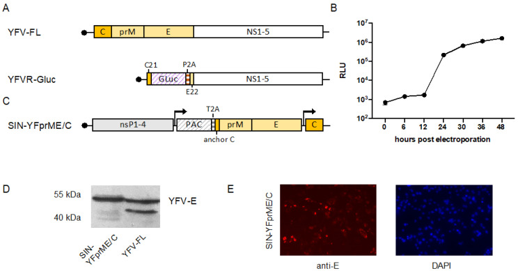
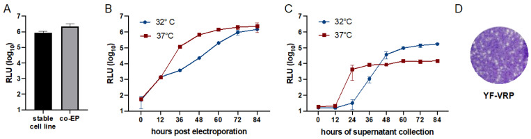
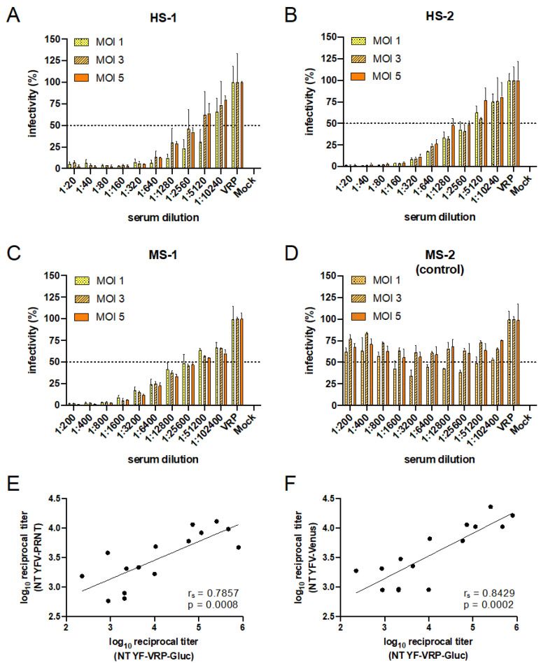
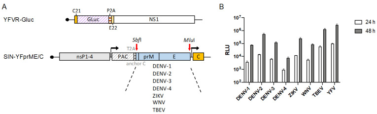
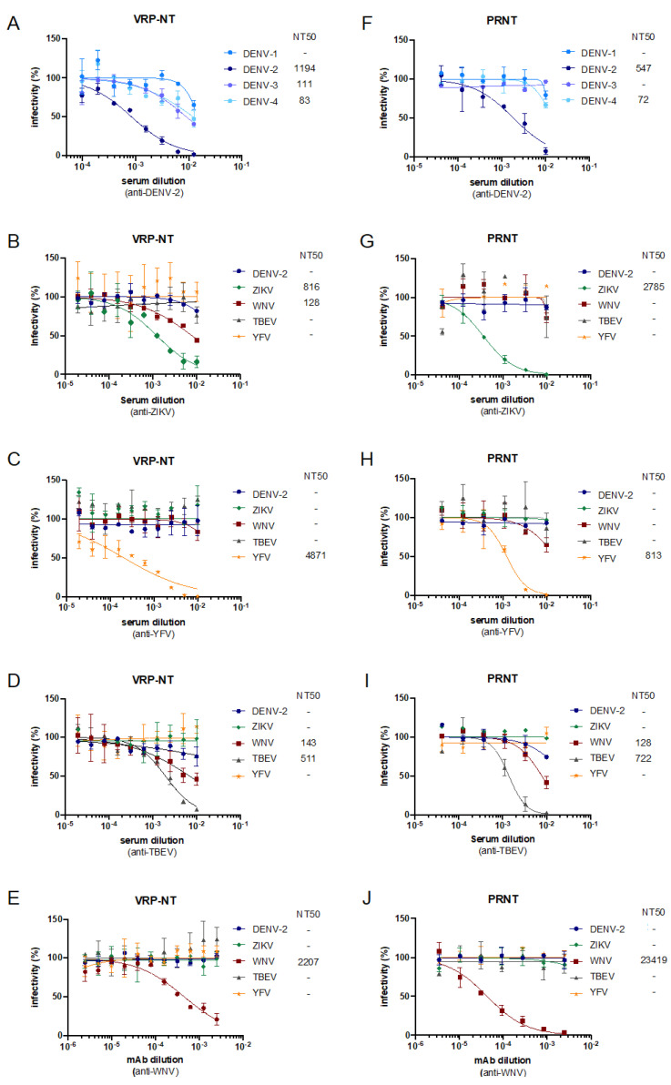
Flavivirus outbreaks require fast and reliable diagnostics that can be easily adapted to newly emerging and re-emerging flaviviruses. Due to the serological cross-reactivity among flavivirus antibodies, neutralization tests (NT) are considered the gold standard for sero-diagnostics. Here, we first established wild-type single-round infectious virus replicon particles (VRPs) by packaging a yellow fever virus (YFV) replicon expressing Gaussia luciferase (Gluc) with YFV structural proteins in trans using a double subgenomic Sindbis virus (SINV) replicon. The latter expressed the YFV envelope proteins prME via the first SINV subgenomic promoter and the capsid protein via a second subgenomic SINV promoter. VRPs were produced upon co-electroporation of replicon and packaging RNA. Introduction of single restriction enzyme sites in the packaging construct flanking the prME sequence easily allowed to exchange the prME moiety resulting in chimeric VRPs that have the surface proteins of other flaviviruses including dengue virus 1--4, Zika virus, West Nile virus, and tick-borne encephalitis virus. Besides comparing the YF-VRP based NT assay to a YF reporter virus NT assay, we analyzed the neutralization efficiencies of different human anti-flavivirus sera or a monoclonal antibody against all established VRPs. The assays were performed in a 96-well high-throughput format setting with Gluc as readout in comparison to classical plaque reduction NTs indicating that the VRP-based NT assays are suitable for high-throughput analyses of neutralizing flavivirus antibodies.

Keywords: diagnostics; flavivirus; high-throughput; neutralization assay; virus replicon particles.


Conflict of interest statement

The authors declare no conflict of interest.

Figures

Figure 1
Figure 1
Scheme and characterization of components for yellow fever virus replicon particle (YF-VRP) production. (A) Schematic presentation of the YFV replicon expressing Gaussia luciferase (YFVR-Gluc) in comparison to the YFV full-length genome (YFV-FL). Lines indicate non-translated regions with the solid black dots at each 5′ end representing the cap structure. Boxes represent translated regions, with solid-colored boxes indicating the YFV structural proteins, the open box the YFV nonstructural proteins, and hatched boxes the Gluc marker or porcine teschovirus-1 derived autocatalytic peptide 2A (P2A), respectively. C21 represents the first 21 amino acids of the capsid protein and E22 the last 22 amino acids of the envelope protein, which serve as signal sequence for the following NS1 protein. (B) Kinetics of Gluc secretion after electroporation of the YFV replicon expressing Gluc. RNA transcribed in vitro from the replicon was electroporated into BHK cells and Gluc secreted into the supernatant was measured at the indicated time points (RLU: relative light units). (C) Scheme of the double subgenomic noncytopathogenic SINV replicon-based packaging construct expressing the YFV structural proteins (SIN-YFprME/C). Hatched boxes indicate the puromycin N-acetyl-transferase (PAC) or the Thosea asigna virus 2A self-cleavage peptide (T2A), respectively. Other symbols used as described under A. Arrows indicate the positions of the SINV subgenomic promoters. (D) Western blot analyses of YFV E in packaging cell line stably expressing YFprME/C. A lysate of cells infected with YFV served as a positive control. Detection was performed with a rabbit anti-YFV-E antiserum (E) Immunofluorescence analysis of the stable packaging cell line. Indirect immunofluorescence was performed using a pan-flavi antibody recognizing the envelope protein. Nuclei were stained using DAPI.
Figure 2
Figure 2
Optimization of VRP production. (A) In vitro synthesized YFV Gluc replicon RNA was either electroporated into BHK cells stably expressing the flavivirus structural proteins or co-electroporated with in vitro transcribed RNA derived from the packaging construct. Equal amounts of supernatant harvested 72 h after electroporation were used to infect naïve BHK cells. At 24 h after infection, Gluc activity released into the supernatant was measured. (B) In vitro transcribed YFV Gluc replicon RNA was co-electroporated with in vitro transcribed RNA from the packaging construct and cells were incubated at either 32 °C or 37 °C. At the indicated time points, supernatants were harvested and Gluc activity was measured. (C) Equal volumes of supernatants harvested at different time points from the different incubation temperatures in (B) were used to infect fresh BHK cells. At 24 h post-infection, Gluc activity was measured in the supernatant. (D) Plaque formation after titration of YF-VRPs on BHK cells stably expressing YFV structural proteins. After infection, cells were overlaid with agarose and incubated at 37 °C. After three days, cells were fixed and crystal violet staining was performed.
Figure 3
Figure 3
Validation of YF-VRP-Gluc based NT assay. (A,B) NT assays using sera from YFV vaccinated humans were performed in 96-well plates. Human sera were serially diluted and preincubated with VRPs corresponding to an MOI of 1, 3, or 5. BHK cells were infected with the preincubated samples and at 24 h post-infection readout via Gluc secreted into the supernatant was performed. For bars labeled with VRP, cells were infected with the appropriate amount of VRPs not preincubated with serum. The % infectivity was normalized to VRP infection without serum incubations. Data represent mean ± SD of experiments performed in duplicate. (C,D) YFV VRP-Gluc NT assays using an anti-YFV mouse serum (C) or a mouse control serum (D) were performed as described for (A,B). (E) Correlation of NT50 titers obtained via YF-VRP-Gluc NT assay (MOI 5) and NT50 titers obtained via PRNT assay. (F) Correlation of NT50 titers obtained via YF-VRP-Gluc NT assay (MOI 5) and NT50 titers obtained using a full-length YFV-17D-Venus reporter virus (MOI 1). (E,F) Depicted values are derived from duplicates. The lines indicate the linear regressions. rs: Spearman correlation coefficient.
Figure 4
Figure 4
Production of chimeric VRPs. (A) Schematic presentation of co-electroporated RNAs used to produce chimeric VRPs, in which a YFV-Gluc replicon is encapsidated with envelope proteins of other flaviviruses. Different packaging RNAs based on a double subgenomic SINV replicon were used, each expressing the Ca-prM-E sequences of the indicated viruses and the YFV capsid protein. See also Figure 1 for further explanations. (B) Equal volumes of supernatant harvested after co-electroporation were used to infect fresh BHK cells. Gluc activity was measured in the supernatant at the indicated time points after infection. Data represent means ± SD of experiments performed in duplicate.
Figure 5
Figure 5
Comparison of VRP-based NT assays to plaque reduction NT assays. (AE) Neutralization curves for VRP-based NT assays or (FJ) neutralization curves for PRNT-based NT assays using the indicated human anti-flavivirus sera or an anti-WNV mAb. VRP-based NT assays (MOI 1) were performed using two-fold serum dilutions and PRNT assays were done using three-fold serum dilutions. After preincubation of sera/mAb with VRPs/virus, BHK cells were infected with the preincubated samples. At 48 h post infection readout via Gluc secreted into the supernatant was performed. Normalized percentage neutralization values are plotted against the serum dilution factors. Inferred NT50 titers are depicted as the reciprocal of the serum dilution conferring 50% inhibition of VRP or virus infection. Data represent means ± SD of experiments performed in duplicate.

Similar articles

Cited by

References

    1. Pierson T.C., Diamond M.S. The continued threat of emerging flaviviruses. Nat. Microbiol. 2020;5:796–812. doi: 10.1038/s41564-020-0714-0. - DOI - PMC - PubMed
    1. Frierson J.G. The yellow fever vaccine: A history. Yale J. Biol. Med. 2010;83:77–85. - PMC - PubMed
    1. Silva N.I.O., Sacchetto L., de Rezende I.M., Trindade G.S., LaBeaud A.D., de Thoisy B., Drumond B.P. Recent sylvatic yellow fever virus transmission in Brazil: The news from an old disease. Virol. J. 2020;17:9. doi: 10.1186/s12985-019-1277-7. - DOI - PMC - PubMed
    1. de Oliveira Figueiredo P., Stoffella-Dutra A.G., Barbosa Costa G., Silva de Oliveira J., Dourado Amaral C., Duarte Santos J., Soares Rocha K.L., Araújo Júnior J.P., Lacerda Nogueira M., Zazá Borges M.A., et al. Re-Emergence of Yellow Fever in Brazil during 2016–2019: Challenges, Lessons Learned, and Perspectives. Viruses. 2020;12:1233. doi: 10.3390/v12111233. - DOI - PMC - PubMed
    1. WHO Yellow Fever—Brazil. [(accessed on 22 December 2021)]. Available online: https://www.who.int/emergencies/disease-outbreak-news/item/11-february-2....

Publication types

MeSH terms