. 2022 Feb 9;14(2):344.
doi: 10.3390/v14020344.

Elucidation of the Mechanism of Host NMD Suppression by HTLV-1 Rex: Dissection of Rex to Identify the NMD Inhibitory Domain

Affiliations

Elucidation of the Mechanism of Host NMD Suppression by HTLV-1 Rex: Dissection of Rex to Identify the NMD Inhibitory Domain

Kazumi Nakano et al. Viruses. .

Abstract

The human retrovirus human T-cell leukemia virus type I (HTLV-1) infects human T cells by vertical transmission from mother to child through breast milk or horizontal transmission through blood transfusion or sexual contact. Approximately 5% of infected individuals develop adult T-cell leukemia/lymphoma (ATL) with a poor prognosis, while 95% of infected individuals remain asymptomatic for the rest of their lives, during which time the infected cells maintain a stable immortalized latent state in the body. It is not known why such a long latent state is maintained. We hypothesize that the role of functional proteins of HTLV-1 during early infection influences the phenotype of infected cells in latency. In eukaryotic cells, a mRNA quality control mechanism called nonsense-mediated mRNA decay (NMD) functions not only to eliminate abnormal mRNAs with nonsense codons but also to target virus-derived RNAs. We have reported that HTLV-1 genomic RNA is a potential target of NMD, and that Rex suppresses NMD and stabilizes viral RNA against it. In this study, we aimed to elucidate the molecular mechanism of NMD suppression by Rex using various Rex mutant proteins. We found that region X (aa20-57) of Rex, the function of which has not been clarified, is required for NMD repression. We showed that Rex binds to Upf1, which is the host key regulator to detect abnormal mRNA and initiate NMD, through this region. Rex also interacts with SMG5 and SMG7, which play essential roles for the completion of the NMD pathway. Moreover, Rex selectively binds to Upf3B, which is involved in the normal NMD complex, and replaces it with a less active form, Upf3A, to reduce NMD activity. These results revealed that Rex invades the NMD cascade from its initiation to completion and suppresses host NMD activity to protect the viral genomic mRNA.

Keywords: HTLV-1 rex; NMD inhibition; SMG5/7; Upf1; Upf3; viral RNA.


Conflict of interest statement

The authors declare no conflict of interest.

Figures
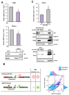
Figure 1
Figure 1
Rex inhibits NMD in T cells. (A). NMD activity was measured in CEM or Molt-4 cells constantly expressing Rex. The results showed a significant decrease in NMD activity in Rex-expressing cells of both cell lines (n = 6, mean ± SD, * p < 0.05). The image below shows Rex expression in Rex(+/-)-Molt-4 cells as detected by Western blotting. The whole-cell lysate of HTLV-1-infected immortalized cell line MT-2 is used as the positive control in Western blotting. (B). NMD-activity reporters (mCherry-WT (red) and EGFP-PTC (green)) were introduced into Jurkat, and the expression levels of mCherry and EGFP in the presence and absence of Rex were detected by flow cytometry (left figure). The results showed that the percentage of cells expressing EGFP increased in Rex-expressing cells compared to Mock cells (right panel), indicating that EGFP-β-globin (PTC) mRNA is stabilized in the presence of Rex. (C). Wild-type HTLV-1 infectious clone, pFL-MT2, or Rex-deficient clone, Rex(-)-pFL-MT2, were transfected into sHeLa cells stably expressing Renilla-WT and Firefly-PTC and evaluated for NMD activity. The results showed that NMD suppression was abolished in Rex (-) clone-transfected cells (top graph). In the cells with Rex(-)-pFL-MT2, the expression of Tax was upregulated and the expression of viral structural proteins such as Gag-p53 and Env-gp24 was markedly reduced (bottom image) (n = 6, mean ± SD, * p < 0.05).
Figure 2
Figure 2
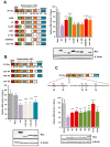
N-terminal region of Rex plays important roles in NMD inhibition. (A). We generated seven domain-deficient mutants of Rex (left panel) and compared the degree of NMD repression with WT-Rex using the NMD activity reporter assay system in HeLa cells. As shown in the graph on the right, NMD repression was significantly reduced when the ARM region, X region or both multimerization domains were deleted. Western blotting below shows the expression levels of each Rex mutant (n = 6, mean ± SD, *** p < 0.001, compared with WT-Rex). (B). For the C-terminal side, where the stabilizing domain of Rex is located, we also generated three types of deletion mutants (upper panel) and compared the degree of NMD repression with WT-Rex using the NMD activity reporter assay system in HeLa cells. As shown in the lower graph, there was no significant change in NMD repression in any of the mutants compared to WT-Rex. Western blotting below shows the expression levels of each Rex mutant (n = 6, mean ± SD, *** p < 0.001, compared with Mock). (C). We generated five point mutants in the ARM and X regions of Rex (upper panel) and compared the degree of NMD repression with WT-Rex using the NMD activity reporter assay system in HeLa cells. As shown in the lower graph, the degree of NMD suppression was significantly reduced in all mutants compared to WT-Rex. The reduction in NMD repression was particularly pronounced for both M1 and M2 mutants. Western blotting below shows the expression levels of each Rex mutant (n = 6, mean ± SD, ** p < 0.01; *** p < 0.001, compared with WT-Rex).
Figure 3
Figure 3
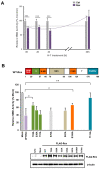
Phosphorylation status of Rex is related to its NMD inhibitory function. (A). Time course changes in the ability of Rex to inhibit NMD after treatment with the PKC inhibitor H-7 were examined using the NMD activity reporter assay system in HeLa cells. The results showed that Rex significantly inhibited NMD activity up to 4 h after H-7 treatment compared with the control but lost its ability to inhibit NMD after 20 h (n = 6, mean ± SD, *** p < 0.001). (B). Each mutant of seven phosphorylation sites of Rex was prepared and the NMD inhibitory ability was compared with that of WT-Rex using the NMD activity reporter assay system in HeLa cells. The results showed that the phosphorylation-deficient mutants at T22 in the X region, S106 in the C-terminal multimerization domain and T174 in the stability domain showed significantly reduced NMD repression. Western blotting below shows the expression levels of each Rex mutant (n = 6, mean ± SD, * p < 0.05; ** p < 0.01).
Figure 4
Figure 4
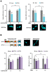
Rex needs to be nuclear-exported to inhibit NMD. (A). Treatment with Leptomycin B, an inhibitor of the nuclear export protein CRM1, significantly reduced the ability of Rex to inhibit NMD (left panel, n = 6, mean ± SD, *** p < 0.001). In contrast, overexpression of CRM1 significantly enhanced the ability of Rex to inhibit NMD (right panel, n = 6, mean ± SD, * p < 0.05; *** p < 0.001). The confocal laser microscopy images below show the subcellular localization of ECFP-Rex in HeLa cells subjected to each treatment. The scale bar indicates 10 μm (B). The data of the p21Rex mutant, which lacks NLS and thus localizes only in the cytoplasm (upper panel, The scale bar indicates 10 μm.), are added to the graphs of WT(p27)-Rex to compare the NMD-suppressive ability of WT(p27)-Rex with that of p21Rex under Leptomycin B treatment or CRM1 overexpression. Upon Leptomycin B treatment, only WT-Rex showed a significant decrease in NMD repression (left panel, n = 6, mean ± SD, ** p < 0.01; *** p < 0.001). When CRM1 was overexpressed, WT-Rex showed a significant enhancement of NMD repression, whereas p21Rex showed no change in NMD repression (right panel, n = 6, mean ± SD, * p < 0.05; *** p < 0.001).
Figure 5
Figure 5
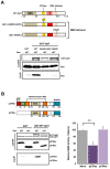
Only WT(p27)-Rex interacts with WT-Upf1. (A). Hyperphosphorylated and RNA helicase-deficient Upf1 mutants were generated (upper panel) and their interaction with WT-Rex was examined by GST pulldown assay in GST-Upf1 and FLAG-Rex-expressing HEK293FT cells. The results showed that Rex interacted only with WT-Upf1 and not with the NMD-deficient Upf1 mutants (lower panel). (B). The interaction of WT-Rex and p21Rex with WT-Upf1 was examined by GST pulldown assay in GST-Upf1 and FLAG-Rex-expressing HEK293FT cells. The results showed that only WT-Rex interacted with Upf1 (bottom left), which was correlated with NMD inhibitory capacity measured by the NMD activity reporter assay system in HeLa cells (bottom right) (n = 6, mean ± SD, ** p < 0.01).
Figure 6
Figure 6
Rex interacts with Upf1 in the cytoplasm and in the p-body. (A). Confocal laser microscopy images of ECFP-Rex, EYFP-Upf1, and mCherry-Dcp2 in HeLa cells, where Dcp2 is a p-body marker. The white arrowhead indicates the site of the p-body. The scale bar indicates 10 μm. (B). The strength of the interaction between Rex and Upf1 in the cytoplasm, the p-body and the nucleus was analyzed by FRET. The left panel shows an example of images before and after acceptor photobleaching by confocal laser microscopy in HEK293FT cells co-expressing ECFP-Rex and EYFP-Upf1. The dotted white circles indicate regions where photobleaching was applied (Cy = cytoplasm, P = p-body, and Nu = nucleus, The scale bar indicates 10 μm.). The graph on the right shows the FRET efficiency (%). Significantly higher FRET efficiency was detected in the positive control (tandem ECFP + EYFP) than in the negative controls (ECFP and EYFP, ECFP and EYFP-Upf1, ECFP-Rex and EYFP), demonstrating that FRET efficiency is correctly measured in this method. The FRET efficiency between ECFP-Rex and EYFP-Upf1 was significantly higher in the cytoplasm and the p-body, indicating that Rex and Upf1 interact at these intracellular sites (n = 6–10, mean ± SD, *** p < 0.001).
Figure 7
Figure 7
Rex interacts with Upf1 and SMG5/7 and contributes to the substitution of Upf3B and Upf3A. (A). NMD regulatory complex proteins interacting with Rex in HEK293FT cells were investigated by GST-Rex coimmunoprecipitation assay. The results showed that Rex interacts not only with Upf1 but also with SMG5, SMG7 and Upf3B. In contrast, Rex did not interact with Upf3A, the isoform of Upf3B. (B). Amount of Upf3B which binds to Upf2 in the NMD complex in the presence or absence of Rex detected by coimmunoprecipitation assay of GST-Upf2 with His-Upf3B and HA-Upf3A in HEK293FT cells. The amount of Upf3B interacting with Upf2 was reduced in the presence of Rex, and instead the interaction with Upf3A, the less active form of Upf3, was increased.
Figure 8
Figure 8
Possible model of Rex-NMD complex interaction and new questions. NMD progresses in a cascade as shown in (1–6). (1): Our results suggest that Rex may inhibit NMD by intervening from the beginning to the end of this cascade by interacting with the NMD key regulator Upf1 and may have some effect on its activity (Baloon-1). (2): Rex interacts only with Upf3B, suggesting that Rex substitutes Upf3A for Upf3B in the NMD complex, resulting in the inclusion of less active Upf3A in the NMD complex, thereby suppressing NMD activity (Baloon-2). (4,5): The mRNA decay factors, the SMG5/7 complex, are recruited to phosphorylated Upf1. The SMG5/7 complex recruits the decapping complex (DCP2/DCP1a) and the deadenylation complex CCR4–NOT to enhance decapping and deadenylation of the NMD target mRNA. Additionally, the SMG5/7 complex recruits protein phosphatase 2A (PP2A) for dephosphorylation of Upf1. Since dephosphorylation of Upf1 is essential for NMD completion, Rex may influence the dephosphorylation pathway of Upf1 via SMG5/7 interaction, and thus may influence NMD completion (Baloon-3).

Similar articles

Cited by

References

    1. Poiesz B.J., Ruscetti F.W., Gazdar A.F., Bunn P.A., Minna J.D., Gallo R.C. Detection and isolation of type C retrovirus particles from fresh and cultured lymphocytes of a patient with cutaneous T-cell lymphoma. Proc. Natl. Acad. Sci. USA. 1980;77:7415–7419. doi: 10.1073/pnas.77.12.7415. - DOI - PMC - PubMed
    1. Poiesz B.J., Ruscetti F.W., Reitz M.S., Kalyanaraman V.S., Gallo R.C. Discovery of new type C retrovirus (HTLV-1) Nature. 1981;294:268–271. doi: 10.1038/294268a0. - DOI - PubMed
    1. Yoshida M., Miyoshi I., Yorio H. Isolation and characterization of retrovirus from cell lines of human adult T-cell leukemia and its implications in the disease. Proc. Natl. Acad. Sci. USA. 1982;79:2031–2035. doi: 10.1073/pnas.79.6.2031. - DOI - PMC - PubMed
    1. Takatsuki K. Discovery of adult T-cell leukemia. Retrovirology. 2005;3:16. doi: 10.1186/1742-4690-2-16. - DOI - PMC - PubMed
    1. Gallo R.C. The discovery of the first human retrovirus: HTLV-1 and HTLV-2. Retrovirology. 2005;7:17. doi: 10.1186/1742-4690-2-17. - DOI - PMC - PubMed

Publication types

MeSH terms

LinkOut - more resources