. 2022 Feb 9;14(2):359.
doi: 10.3390/v14020359.

Encephalomyocarditis Virus 2A Protein Inhibited Apoptosis by Interaction with Annexin A2 through JNK/c-Jun Pathway

Affiliations

Encephalomyocarditis Virus 2A Protein Inhibited Apoptosis by Interaction with Annexin A2 through JNK/c-Jun Pathway

Ruochan Han et al. Viruses. .

Abstract

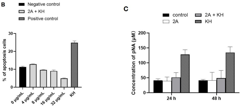
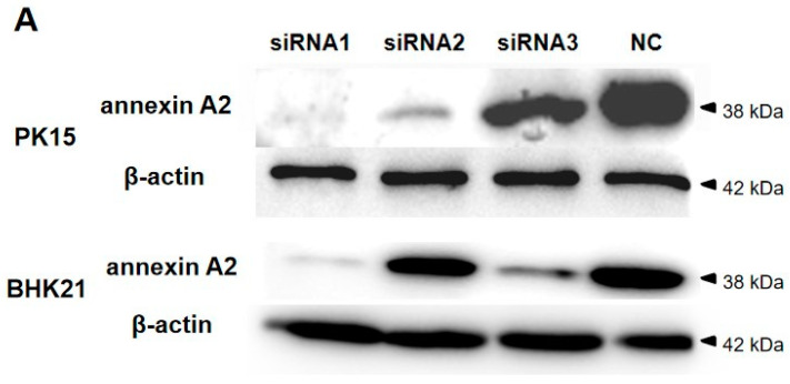
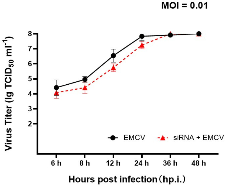
Encephalomyocarditis virus can cause myocarditis and encephalitis in pigs and other mammals, thus posing a potential threat to public health safety. The 2A protein is an important virulence factor of EMCV. Previous studies have shown that the 2A protein may be related to the inhibition of apoptosis by virus, but its specific molecular mechanism is not clear. In this study, the 2A protein was expressed in Escherichia coli in order to find interacting cell proteins. A pull down assay, coupled with mass spectrometry, revealed that the 2A protein possibly interacted with annexin A2. Co-immunoprecipitation assays and confocal imaging analysis further demonstrated that the 2A protein interacted with annexin A2 in cells. In reducing the expression of annexin A2 by siRNA, the ability of the 2A protein to inhibit apoptosis was weakened and the proliferation of EMCV was slowed down. These results suggest that annexin A2 is closely related to the inhibition of apoptosis by 2A. Furthermore, both RT-PCR and western blot results showed that the 2A protein requires annexin A2 interaction to inhibit apoptosis via JNK/c-Jun pathway. Taken together, our data indicate that the 2A protein inhibits apoptosis by interacting with annexin A2 via the JNK/c-Jun pathway. These findings provide insight into the molecular pathogenesis underlying EMCV infection.

Keywords: 2A; JNK/c-Jun pathway; annexin A2; apoptosis; encephalomyocarditis virus.


Conflict of interest statement

The author declares that there are no conflict of interest.

Figures

Figure 1
Figure 1
2A protein inhibits apoptosis in PK15 cells. (A) PK15 cells were treated with KH and different concentrations of pcDNA3.1-2A for 24 h and analysed by flow cytometry. (B) Percentages of Annexin-V-FITC and PI-positive cells from gated cells. (C) The changes of caspase 3 in PK15 cells induced by 8 μg mL−1 pcDNA3.1-2A for 24 h or 48 h. The data are presented as the mean values and standard deviations from three independent experiments.
Figure 1
Figure 1
2A protein inhibits apoptosis in PK15 cells. (A) PK15 cells were treated with KH and different concentrations of pcDNA3.1-2A for 24 h and analysed by flow cytometry. (B) Percentages of Annexin-V-FITC and PI-positive cells from gated cells. (C) The changes of caspase 3 in PK15 cells induced by 8 μg mL−1 pcDNA3.1-2A for 24 h or 48 h. The data are presented as the mean values and standard deviations from three independent experiments.
Figure 2
Figure 2
Identification of 2A-interacting protein by His pull-down assay. Lane 1, proteins of PK15 cells; lane 2, purified 2A protein; lane 3, the specific proteins that may interact with 2A protein by pull down assay. 2A protein produced in E. coli BL21 (DE3) was used for the His pull-down assay.
Figure 3
Figure 3
Identification of the interaction between the 2A protein and annexin A2. (A) The results of Co-IP. The plasmid (+) or empty vectors (−) were transfected into PK15 cells, and the whole-cell lysates obtained at 48 h post-transfection were immunoprecipitated (IP) with anti-HA pcAb. Then, proteins separated by SDS-PAGE were detected by immunoblotting with the indicated antibodies. The proteins were labeled on the right of the band. (B) Colocalization of the 2A protein with endogenous annexin A2 which is in the membranes. The PK15 cells were stimulated with 4 μg mL−1 pEGFP-2A. Cells were fixed 48 h after plasmids transfection and examined by confocal microscopy to detect 2A (green) and membranes (red). The membranes were labeled with DiI (red) stain. The position of the nucleus is indicated by DAPI (blue) staining in the merged image. Bars = 8 μm. (C) Colocalization of 2A protein and annexin A2. PK15 cells were co-transfected with pEGFP-2A and pDsRed-ANXA2. Cells were fixed at 48 hpt and examined by confocal microscopy to detect 2A (green) and annexin A2 (red). The position of the nucleus is indicated by DAPI (blue) staining in the merged image. Bars = 10 μm. Experiments were performed at least three times.
Figure 3
Figure 3
Identification of the interaction between the 2A protein and annexin A2. (A) The results of Co-IP. The plasmid (+) or empty vectors (−) were transfected into PK15 cells, and the whole-cell lysates obtained at 48 h post-transfection were immunoprecipitated (IP) with anti-HA pcAb. Then, proteins separated by SDS-PAGE were detected by immunoblotting with the indicated antibodies. The proteins were labeled on the right of the band. (B) Colocalization of the 2A protein with endogenous annexin A2 which is in the membranes. The PK15 cells were stimulated with 4 μg mL−1 pEGFP-2A. Cells were fixed 48 h after plasmids transfection and examined by confocal microscopy to detect 2A (green) and membranes (red). The membranes were labeled with DiI (red) stain. The position of the nucleus is indicated by DAPI (blue) staining in the merged image. Bars = 8 μm. (C) Colocalization of 2A protein and annexin A2. PK15 cells were co-transfected with pEGFP-2A and pDsRed-ANXA2. Cells were fixed at 48 hpt and examined by confocal microscopy to detect 2A (green) and annexin A2 (red). The position of the nucleus is indicated by DAPI (blue) staining in the merged image. Bars = 10 μm. Experiments were performed at least three times.
Figure 4
Figure 4
Reducing of annexin A2 attenuated the apoptosis inhibited by the 2A protein. (A) Reducing of annexin A2 expression by siRNA treatment. PK15 and BHK21 cells transfected with negative siRNA (NC) or treated with siRNAs separately against annexin A2 (100 pM siRNA1, siRNA2, or siRNA3) were harvested at 48 hpt. Endogenous annexin A2 of cells was detected by immunoblotting with anti-annexin A2 Abs. (B) The apoptosis levels of PK15 and BHK21 cells were detected by flow cytometry. Cells treated with 100 pM siRNA1 for 24 h were transfected with the indicated concentrations of pcDNA3.1-2A and added KH for 24 hpt. (C) Percentages of Annexin-V-FITC and PI positive cells from gated cells. The data are presented as the mean values and standard deviations from three independent experiments.
Figure 4
Figure 4
Reducing of annexin A2 attenuated the apoptosis inhibited by the 2A protein. (A) Reducing of annexin A2 expression by siRNA treatment. PK15 and BHK21 cells transfected with negative siRNA (NC) or treated with siRNAs separately against annexin A2 (100 pM siRNA1, siRNA2, or siRNA3) were harvested at 48 hpt. Endogenous annexin A2 of cells was detected by immunoblotting with anti-annexin A2 Abs. (B) The apoptosis levels of PK15 and BHK21 cells were detected by flow cytometry. Cells treated with 100 pM siRNA1 for 24 h were transfected with the indicated concentrations of pcDNA3.1-2A and added KH for 24 hpt. (C) Percentages of Annexin-V-FITC and PI positive cells from gated cells. The data are presented as the mean values and standard deviations from three independent experiments.
Figure 5
Figure 5
Reducing of annexin A2 increased apoptosis in the infection of EMCV. (A) The apoptosis levels of PK15 and BHK21 cells were detected by flow cytometry. PK15 and BHK21 cells treated with 100 pM siRNA1 were infected with MOI of 0.01 with EMCV-HB10 for 24 hpt. (B) Percentages of Annexin-V-FITC and PI positive cells from gated cells. The data are presented as the mean values and standard deviations from three independent experiments.
Figure 5
Figure 5
Reducing of annexin A2 increased apoptosis in the infection of EMCV. (A) The apoptosis levels of PK15 and BHK21 cells were detected by flow cytometry. PK15 and BHK21 cells treated with 100 pM siRNA1 were infected with MOI of 0.01 with EMCV-HB10 for 24 hpt. (B) Percentages of Annexin-V-FITC and PI positive cells from gated cells. The data are presented as the mean values and standard deviations from three independent experiments.
Figure 6
Figure 6
Effect of annexin A2 on EMCV proliferation. One-step growth curves of EMCV-HB10 in BHK21 (●) and BHK21 reducing annexin A2 (▲). An MOI of 0.01 was used for infection, supernatant and cells were harvested at each indicated time and freeze-thawed together for viral titration, as described in Methods.
Figure 7
Figure 7
The 2A protein inhibits apoptosis by interacting with annexin A2 via JNK/c-Jun pathway in BHK21 cells. (A) BHK21 cells were treated with annexin A2 siRNA1 alone, or together with pcDNA3.1-2A or alone with pcDNA3.1-2A were harvested at 48 hpt. The mRNA expression of caspase 3/8/9 were detected by Real-time RT-PCR analysis. GAPDH was used as an internal control. (B) Cells were treated in the same way as (A), the mRNA expression of p53 and c-Jun were detected by RT-PCR analysis. (C) Cells were treated in the same way as (A), the expression of caspase 3/8/9, p53, p-JNK and p-c-Jun were detected by Western blot. Anti-β-actin antibody was used to confirm comparable loading. (D) BHK21 cells were treated with different concentrations of pcDNA3.1-2A for 24 h, and then the mRNA expression of annexin A2 and c-Jun were detected by RT-PCR analysis. (E) BHK21 cells were treated as (D) for 48 h, and then the expression of 2A, annexin A2, p-JNK and p-c-Jun were detected by Western blot. (F,G) BHK-21 cells were infected at an MOI of 0.01 with EMCV, and at each indicated time, supernatant and cells were harvested. The mRNA expression of caspase 3/8, p53 and c-Jun were detected by RT-PCR analysis. (H) Cells were treated in the same way as (F), the expression of caspase 8, annexin A2, p53, p-JNK and p-c-Jun were detected by Western blot. The data are presented as the mean values and standard deviations from three independent experiments.
Figure 7
Figure 7
The 2A protein inhibits apoptosis by interacting with annexin A2 via JNK/c-Jun pathway in BHK21 cells. (A) BHK21 cells were treated with annexin A2 siRNA1 alone, or together with pcDNA3.1-2A or alone with pcDNA3.1-2A were harvested at 48 hpt. The mRNA expression of caspase 3/8/9 were detected by Real-time RT-PCR analysis. GAPDH was used as an internal control. (B) Cells were treated in the same way as (A), the mRNA expression of p53 and c-Jun were detected by RT-PCR analysis. (C) Cells were treated in the same way as (A), the expression of caspase 3/8/9, p53, p-JNK and p-c-Jun were detected by Western blot. Anti-β-actin antibody was used to confirm comparable loading. (D) BHK21 cells were treated with different concentrations of pcDNA3.1-2A for 24 h, and then the mRNA expression of annexin A2 and c-Jun were detected by RT-PCR analysis. (E) BHK21 cells were treated as (D) for 48 h, and then the expression of 2A, annexin A2, p-JNK and p-c-Jun were detected by Western blot. (F,G) BHK-21 cells were infected at an MOI of 0.01 with EMCV, and at each indicated time, supernatant and cells were harvested. The mRNA expression of caspase 3/8, p53 and c-Jun were detected by RT-PCR analysis. (H) Cells were treated in the same way as (F), the expression of caspase 8, annexin A2, p53, p-JNK and p-c-Jun were detected by Western blot. The data are presented as the mean values and standard deviations from three independent experiments.

Similar articles

Cited by

References

    1. Carocci M., Bakkali-Kassimi L. The encephalomyocarditis virus. Virulence. 2012;3:351–367. doi: 10.4161/viru.20573. - DOI - PMC - PubMed
    1. Agol V.I., Gmyl A.P. Viral security proteins: Counteracting host defences. Nat. Rev. Microbiol. 2010;8:867–878. doi: 10.1038/nrmicro2452. - DOI - PMC - PubMed
    1. Carocci M., Cordonnier N., Huet H., Romey A., Relmy A., Gorna K., Blaise-Boisseau S., Zientara S., Kassimi L.B. Encephalomyocarditis virus 2A protein is required for viral pathogenesis and inhibition of apoptosis. J. Virol. 2011;85:10741–10754. doi: 10.1128/JVI.00394-11. - DOI - PMC - PubMed
    1. Hajjar K.A., Krishnan S. Annexin II: A mediator of the plasmin/plasminogen activator system. Trends Cardiovasc. Med. 1999;9:128–138. doi: 10.1016/S1050-1738(99)00020-1. - DOI - PubMed
    1. Jolly C., Winfree S., Hansen B., Steele-Mortimer O. The Annexin A2/p11 complex is required for efficient invasion of Salmonella typhimurium in epithelial cells. Cell. Microbiol. 2014;16:64–77. doi: 10.1111/cmi.12180. - DOI - PMC - PubMed

Publication types

MeSH terms

LinkOut - more resources