. 2022 Feb 14;14(2):384.
doi: 10.3390/v14020384.

Phage-Displayed Mimotopes of SARS-CoV-2 Spike Protein Targeted to Authentic and Alternative Cellular Receptors

Affiliations

Phage-Displayed Mimotopes of SARS-CoV-2 Spike Protein Targeted to Authentic and Alternative Cellular Receptors

Valery A Petrenko et al. Viruses. .

Abstract

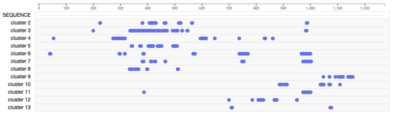
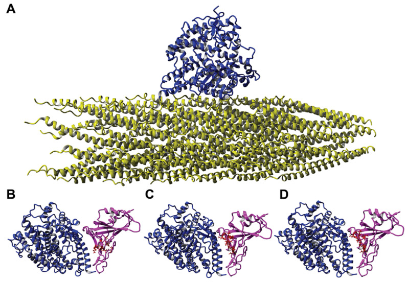
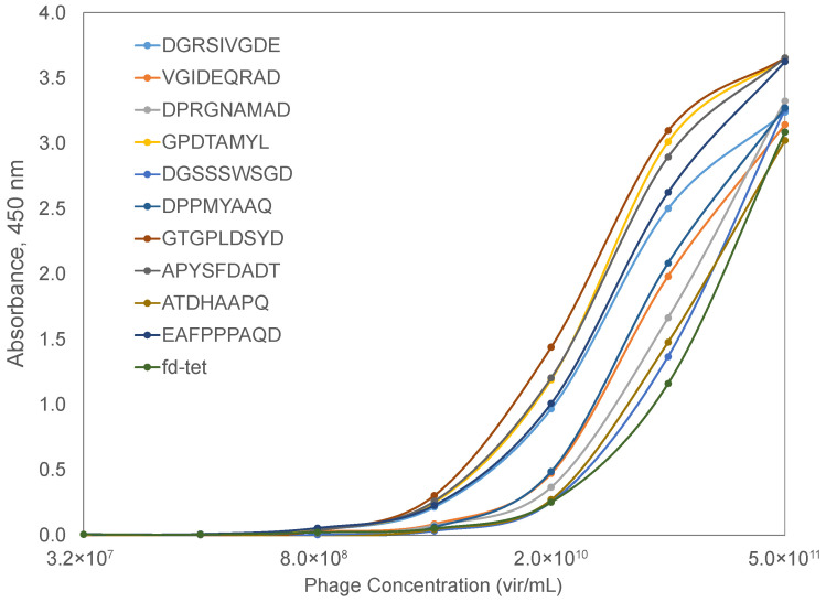
The evolution of the SARS-CoV-2 virus during the COVID-19 pandemic was accompanied by the emergence of new heavily mutated viral variants with increased infectivity and/or resistance to detection by the human immune system. To respond to the urgent need for advanced methods and materials to empower a better understanding of the mechanisms of virus's adaptation to human host cells and to the immuno-resistant human population, we suggested using recombinant filamentous bacteriophages, displaying on their surface foreign peptides termed "mimotopes", which mimic the structure of viral receptor-binding sites on the viral spike protein and can serve as molecular probes in the evaluation of molecular mechanisms of virus infectivity. In opposition to spike-binding antibodies that are commonly used in studying the interaction of the ACE2 receptor with SARS-CoV-2 variants in vitro, phage spike mimotopes targeted to other cellular receptors would allow discovery of their role in viral infection in vivo using cell culture, tissue, organs, or the whole organism. Phage mimotopes of the SARS-CoV-2 Spike S1 protein have been developed using a combination of phage display and molecular mimicry concepts, termed here "phage mimicry", supported by bioinformatics methods. The key elements of the phage mimicry concept include: (1) preparation of a collection of p8-type (landscape) phages, which interact with authentic active receptors of live human cells, presumably mimicking the binding interactions of human coronaviruses such as SARS-CoV-2 and its variants; (2) discovery of closely related amino acid clusters with similar 3D structural motifs on the surface of natural ligands (FGF1 and NRP1), of the model receptor of interest FGFR and the S1 spike protein; and (3) an ELISA analysis of the interaction between candidate phage mimotopes with FGFR3 (a potential alternative receptor) in comparison with ACE2 (the authentic receptor).

Keywords: SARS-CoV-2 virus; alternative receptors; landscape phage; mimotope; molecular mimicry; phage display; spike protein; virus receptors.


Conflict of interest statement

The authors declare no competing financial interest.

Figures

Figure 1
Figure 1
Three-dimensional predicted model of the spike (S) glycoprotein of the SARS-CoV-2 virus composed of (A) two well-defined structural domains (S1 and S2), decorated with (B) 22 N-glycan residues as modeled using 6VSB and 6VXX [38,39]. Monomers of the S protein, composed of polypeptide chains of 1273 amino acids, form homotrimer spikes on the virus surface [18]. Spike protein monomers are composed of three major structural domains: head, stalk, and cytoplasmic tail. The head comprises the N-terminal domain (NTD; yellow) and the receptor-binding domain (RBD; orange), which displays the receptor-binding motif (RBM; cyan) that is responsible for interaction with cell receptors [40]. RBDs in non-activated viral S glycoprotein trimers are present in a hidden “down” conformation. The S glycoprotein is cleaved by host proteases (trypsin and furin) at the site between the S1 and the S2 subunits [41,42]. The S2 domain of the S protein consists of fusion peptide (FP; purple), two heptad-repeat domains (HR1 and HR2; red), a transmembrane domain (TM; gray), and a cytoplasm domain (CP; pink). A second proteolytic site (S2′ site), located within the S2 subdomain, is also cut by type II transmembrane serine protease (TMPRSS2) as well as cathepsin B and L (CatB/L) to enable virus-cell fusion by triggering the dissociation of S1 and the irreversible refolding of S2, a conformational change of the S protein and the fusion of the viral envelope and endosomes [42].
Figure 2
Figure 2
Electron microscopy image of filamentous phage (left) and electron density model (center) of filamentous phage M13 (courtesy of Lee Makowski and Gregory Kishchenko). Blue and red arrows depict the sharp and blunt ends of the phage capsid with attached minor coat proteins pIII/pIV and pVII/pIX, respectively (five copies each). Major coat protein (~2700 copies) forms the tubular capsid around viral single-stranded DNA (scale bar: 100 nm). 3D structure (right) of the complex between phage displaying the peptide EDYSELVSQ (green) with FGFR3 (red). Here, the peptide-displayed phages are designated by the structure of the inserted foreign peptides.
Figure 3
Figure 3
(A) Schematic of viral evolution (adaptation) leading to increased fitness to cellular receptors through diversification of viral functional domains; (B) schematic of SARS-CoV-2 functional domains and epitopes (or their mimetics) fused to p8 proteins and selected from landscape phage libraries through phage mimicry; (C) the phage-displayed peptide (mimotope) contains the same or similar amino acid (AA) residues as amino acid clusters (AA clusters) on the surface of spike protein, and presumably can interact with viral cellular receptors.
Figure 4
Figure 4
Clusters of amino acids identified by PepSurf surface accessible alignments of cell receptor-binding phage mimotopes on the SARS-CoV-2 spike protein using the PDB model 6VYB. Cluster 1 (dark purple), cluster 2 (green), cluster 3 (bright pink), cluster 4 (turquoise), cluster 5 (pink), cluster 6 (dark blue), cluster 7 (purple), cluster 8 (blue), cluster 9 (dark pink), cluster 10 (light blue), cluster 11 (yellow), cluster 12 (orange), and cluster 13 (red).
Figure 5
Figure 5
Amino acid residues corresponding to each identified functional cluster.
Figure 6
Figure 6
Interaction of ACE2 with different ligands. (A) Interaction of a landscape phage displaying the peptide DGRADLSYD on the full-length p8 protein (yellow) with ACE2 (blue) as determined using homology modeling. Here, a segment containing less than 1% of the landscape phage is presented, where the DGRADLSYD peptide is presented as an N-terminal fusion to all copies of the mature p8 major coat protein. Molecular model 6M0J demonstrating the interaction between ACE2 protein (blue) and recombinant SARS-CoV-2 spike RBD (pink) with amino acid clusters corresponding to phage mimotopes. (B) DGRADLSYD; (C) VGIDEQRAD; and (D) DGRSIVGDE, highlighted in red.
Figure 7
Figure 7
ACE2-binding phages displaying SARS-CoV-2 spike RBD-mimotopes as characterized by an indirect ELISA. Serial dilutions of phages were incubated with a bound ACE2 receptor, followed by incubation with a rabbit anti-phage IgG and an HRP-conjugated goat anti-rabbit IgG. Signal was produced using a TMB substrate and endpoint absorbance at 450 nm was measured after a 30-min incubation. Candidate RBD-binding phages were compared to the wildtype fd-tet phage (dark green).
Figure 8
Figure 8
Interaction of fibroblast growth factor receptor 3 (FGFR3) with different ligands: (A) Interaction of a landscape phage displaying the peptide EDYSELVSQ (yellow) on the full-length p8 protein with FGFR3 (cyan), as determined using homology modeling. Here, a segment containing less than 1% of the landscape phage is presented, where the EDYSELVSQ peptide is presented as an N-terminal fusion to all copies of the mature p8 major coat protein. Interaction of FGFR3 (cyan) with (B) FGF1 (gray), (C) NRP1 (magenta), or (D) SARS-CoV-2 spike RBD (blue), with amino acid clusters containing alignments to the EDYSELVSQ mimotope highlighted in red.
Figure 9
Figure 9
Primary structure of the FGFR3 domain. Amino acids involved in the interaction (designated by capital bold letters) between FGF1 (*), NRP1 (^), and RBD (#) were identified using the YASARA Structure and literature data. Promiscuous amino acid residues of FGFR3 involved with the interaction of FGF1, NRP1, and spike RBD are marked with (*^#). Members of AA clusters corresponding to the phage mimotope EDYSELVSQ are marked with (+) and indicated by underlined letters.
Figure 10
Figure 10
FGFR3-binding phages displaying FGF1 mimotopes as characterized by an indirect ELISA. Serial dilutions of phages were incubated with the bound FGFR3 receptor, followed by incubation with a rabbit anti-phage IgG and an HRP-conjugated goat anti-rabbit IgG. Signal was produced using a TMB substrate and endpoint absorbance at 450 nm was measured after a 30-min incubation. Candidate FGFR3-binding phages were compared to the wildtype fd-tet phage (dark green).
Figure 11
Figure 11
Evaluation of receptors interacting with the SARS-CoV-2 S1 spike protein by an indirect ELISA. Recombinant SARS-CoV-2 S1-His protein was incubated with bound ACE2, DPP4, FGFR3, NRP1, or BSA protein in triplicate wells for 1 h at 37 °C. The wells were incubated with a rabbit anti-SARS-CoV-2 spike IgG and an HRP-conjugated goat anti-rabbit IgG for 1 h at 37 °C. Signal was produced using a TMB substrate and endpoint absorbance at 450 nm was measured after a 30-min incubation. Mean endpoint absorbances for recombinant protein receptors were compared to the means of an unrelated BSA control using a Dunnett’s test with statistically significant differences (p < 0.05) indicated with a star.
Figure 12
Figure 12
Primary protein structures of the RBD domain of SARS-CoV-2 (upper line) and SARS-CoV (bottom line) aligned using BLASTP. Amino acid residues involved in the interaction (designated by capital bold letters) between FGFR3 (marked with a *) and ACE2 (marked with a ^) were identified using the YASARA Structure and literature data [97]. Promiscuous AA of the SARS-CoV-2 RBD involved in binding both FGFR3 and ACE2 are marked with *^. Members of AA cluster D442, N448, Y449, S494, Q493, L492, corresponding to phage mimotope EDYSELVSQ, are underlined and marked with (+).

Similar articles

Cited by

References

    1. Millet J.K., Jaimes J.A., Whittaker G.R. Molecular diversity of coronavirus host cell entry receptors. FEMS Microbiol. Rev. 2021;45:fuaa057. doi: 10.1093/femsre/fuaa057. - DOI - PMC - PubMed
    1. Zhou W., Wang W. Fast-spreading SARS-CoV-2 variants: Challenges to and new design strategies of COVID-19 vaccines. Signal Transduct. Target. 2021;6:226. doi: 10.1038/s41392-021-00644-x. - DOI - PMC - PubMed
    1. van Oosterhout C., Hall N., Ly H., Tyler K.M. COVID-19 evolution during the pandemic Implications of new SARS-CoV-2 variants on disease control and public health policies. Virulence. 2021;12:507–508. doi: 10.1080/21505594.2021.1877066. - DOI - PMC - PubMed
    1. Li Q., Wu J., Nie J., Zhang L., Hao H., Liu S., Zhao C., Zhang Q., Liu H., Nie L., et al. The Impact of Mutations in SARS-CoV-2 Spike on Viral Infectivity and Antigenicity. Cell. 2020;182:1284–1294e1289. doi: 10.1016/j.cell.2020.07.012. - DOI - PMC - PubMed
    1. Weisblum Y., Schmidt F., Zhang F., DaSilva J., Poston D., Lorenzi J.C., Muecksch F., Rutkowska M., Hoffmann H.H., Michailidis E., et al. Escape from neutralizing antibodies by SARS-CoV-2 spike protein variants. Elife. 2020;9:e61312. doi: 10.7554/eLife.61312. - DOI - PMC - PubMed

Publication types

MeSH terms

Substances

Supplementary concepts

LinkOut - more resources